Quiz introduction to abdominal x rays. The stomach is in the left upper quadrant and is visible when it is filled with air.
 Film Critique Part 2 Abdomen
6 gas in sigmoid.
Abdominal x ray anatomy. Performing this projection the patient is either supine or upright position as requested by physician. The following questions give you the opportunity to assess and to apply the knowledge skills and attitudes you have learned by working through this e learning module introduction to abdominal x rays. Abdominal organs the parenchymal organs within the abdomen absorb x rays as they pass through the patient and therefore alter the appearance of the radiograph.
This page is dedicated to providing a guide on the approach to interpreting an abdominal x ray. Because of the difference in x ray absorption by air and soft tissues the intestinal structures intestinal air can be differentiated from their surroundings. When an ap projection of abdomen taken in supine it is also called as flat plate abdomen x ray.
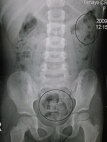
4 gas in colon splenic flexure. We are pleased to provide you with the picture named anatomy on the abdominal x ray. Full assessment includes a check of patient data image quality and checking for artifact and abnormal calcification.
This webpage presents the anatomical structures found on abdominal x ray. 3 gas in stomach. This type of scan is often ordered given that it is fairly quick easy and cheap to obtain and can provide some insight regarding abdominal processes.
A plain x ray of the abdomen can help see the organs and conditions in the belly including intestinal obstruction or perforation. Special projections include a pa prone lateral decubitus upright ap and lateral cross table with the patient supine. This type of scan is also sometimes called a kub kidney ureter and bladder study.
These changes are subtle but with practice you should be able to make out several organs and muscles. But the supine position is preferred for most initial examination of the abdomen. This involves assessment of the bowel gas pattern soft tissue structures and bones.
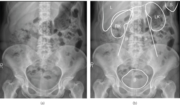
10 gas in cecum 11 iliac crest. Anatomy on the abdominal x ray anatomy on the abdominal x ray in this image you will find right 12 the rib liver gas within the bowel cholecystectomy surgical clip inferior pole of left kidney left sacroiliac joint fat fold in it. The standard abdominal x ray protocol is usually a single anteroposterior projection in supine position.
2 vertebral body th 12. 5 gas in transverse colon. A systematic approach to abdominal x ray interpretation is therefore relatively straightforward.
12 gas in colon hepatic flexure.
 Abdominal X Ray Wikipedia
 Radiology Basics Abdomen Anatomy
 Abdomen Anterior Posterior X Ray View Radiology Radiology
 Radiographic Anatomy Abdomen Ap Supine Medical
 Abdominal X Ray An Approach Summary Radiology
 Radiology Normal Chest X Rays Glass Box
![]() Normal Chest X Ray Anatomy Tutorial Kenhub
Normal Chest X Ray Anatomy Tutorial Kenhub
 Anatomy On The Abdominal X Ray
 X Rays Ct Scans And Mris Orthoinfo Aaos
 Ap Projection Erect Position Abdomen Radtechonduty
 Radiology Basics Abdomen Anatomy
 Abdominal X Ray Startradiology
 Abdomen Radiography Kub Ap Or Pa Projection Radtechonduty
![]() Normal Chest X Ray Anatomy Tutorial Kenhub
Normal Chest X Ray Anatomy Tutorial Kenhub
 Labeled Ap Pelvis Xray Anatomy Male Anatomy Radiology
 X Ray Film Of Dog Anterior View Closed Up In Thorax Standard
 Untitled Document
 Abdominal X Rays
 Radiographs Of The Dog
Abdomen Radiology Key
 Kub X Ray Labeled What Is Abdominal X Ray Or Kub

 Film Critique Part 2 Abdomen
 Learn To Read An X Ray Long Beach Animal Hospital


Posting Komentar
Posting Komentar