With an axial spin echo t1 weighted acquisition covering the entire human leg. The included right hip joint is intact.
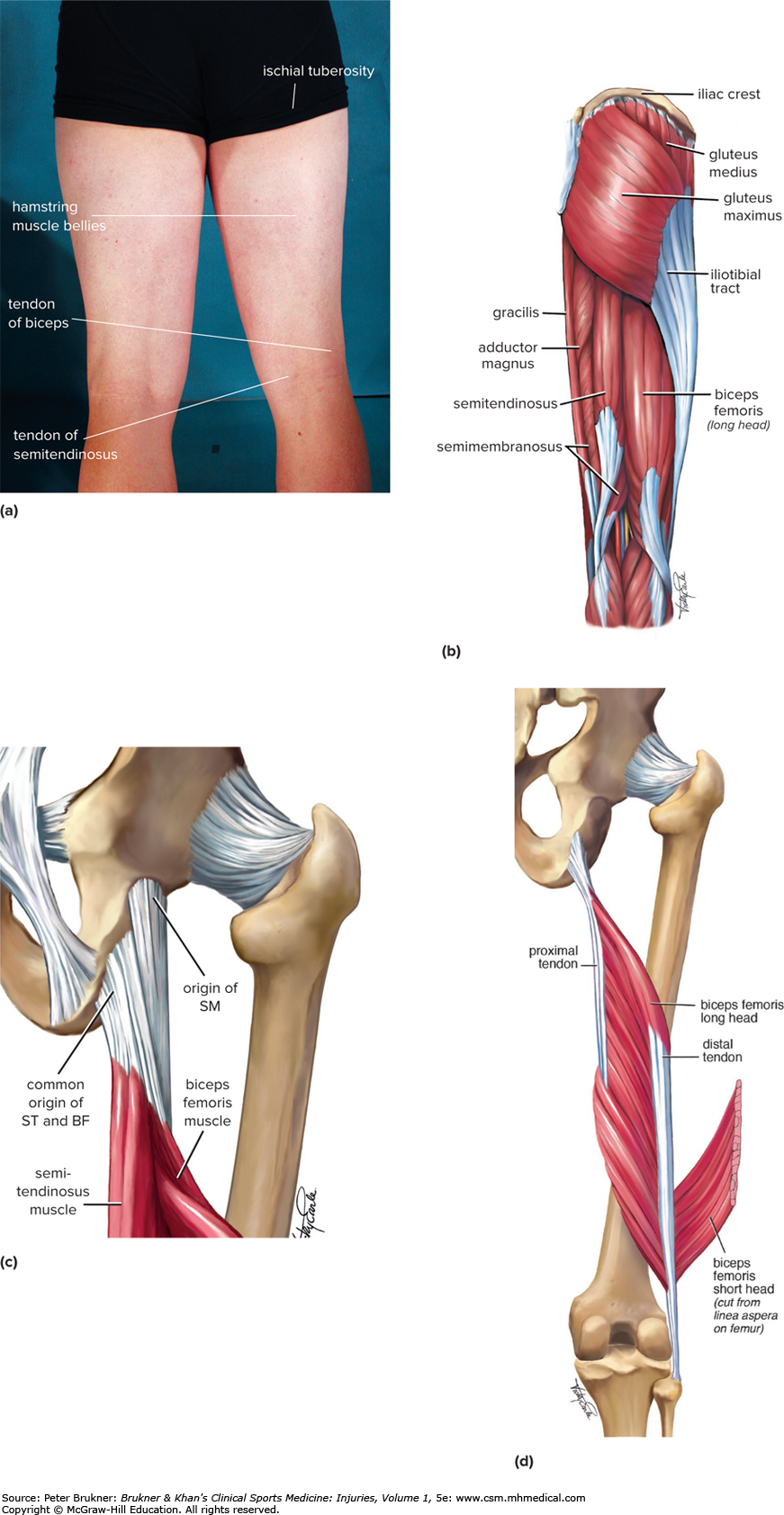 Posterior Thigh Pain Brukner Khan S Clinical Sports
Check for errors and try again.

Mri thigh anatomy. Stanford bone tumor bayesian network issssr msk lectures for residents ocad msk cases from around the world stanford msk mri atlas has served almost 800000 pages to users in over 100 countries. Unable to process the form. The femur shows no definite abnormal marrow signal with intact cortex.
Knee shoulder shoulder arthrogram ankle elbow wrist hip contact. Note that in an anatomical context leg refers to the portion between the knee and ankle joints and not to the entire lower limb. A magnetic resonance imaging mri was performed on a healthy subject.
The muscles are normal in size without signal intensity. Use the mouse to scroll or the arrows. Upper two thirds of the medial margin and proximal margin of the patella medial condyle of the tibia and investing deep fascia of the leg with the tendons of vastus intermedius lateralis and rectus and through the patellar ligament onto the front of the tibial tuberosity.
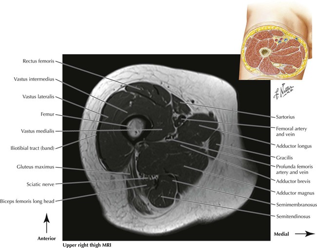
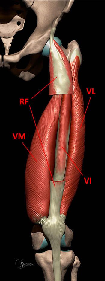
2 vastus medialis intermedius muscles. Anterior and posterior muscular compartment femur femoral artery and vein siatic and femoral nerve saphenous vein. Related posts of thigh muscle anatomy mri eye diagram muscles anatomy.
1 vastus lateralis muscle. Positioning for mri upper legs position the patient in supine position with feet pointing towards the magnet feet first supine position the patient over the spine coil and place the body coils over the thighs anterior superior iliac spine down to knee joints. Eye diagram muscles anatomy 16 photos of the eye diagram muscles anatomy diagram back muscles diagram leg muscles diagram of eye anatomy diagram of muscles in arm diagram of muscles in lower back diagram of muscles in the knee diagram of muscles in the neck human muscles diagram back muscles diagram leg muscles.
Thigh refers to the portion of the lower limb between the hip and knee joints. Anatomy of the thigh.
 Knee Anatomy Mri Knee Coronal Anatomy Free Cross
 Adipose Tissues In The Leg Thigh And The Effect Of

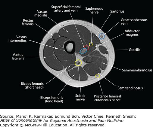 Sonoanatomy Relevant For Ultrasound Guided Lower Extremity
 Mri Thigh Calf Anatomy Normal Anatomy Dr Ahmed Eisawy
 Mri Thigh Upper Legs Planning Mri Hamstring Protocols
 Cross Sectional Anatomy Of The Knee Based On Mri Articular
 Mri Pelvis Anatomy Free Male Pelvis Axial Anatomy
 Mri Of The Thigh Radiology Key
 Figure 4 From Normal Mr Imaging Anatomy Of The Thigh And Leg
 Advanced Body Composition Assessment From Body Mass Index
 Figure 4 From Normal Mr Imaging Anatomy Of The Thigh And Leg
Novel Stochastic Framework For Automatic Segmentation Of
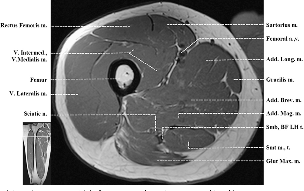 Figure 4 From Normal Mr Imaging Anatomy Of The Thigh And Leg
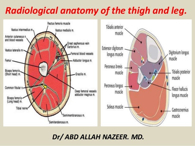 Presentation1 Pptx Radiological Anatomy Of The Thigh And Leg
 Amazon Com Volume Ii Mri Of Sports Hernias Thigh And Hip
 Lower Limbs Radiology Key
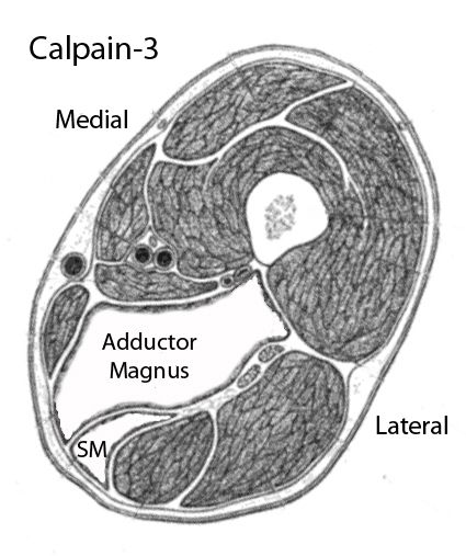
![]() Transverse Mri Scans Of The Qf At 50 Of Femur Length Vi
Transverse Mri Scans Of The Qf At 50 Of Femur Length Vi

Untitled Document
 Eosinophilic Fasciitis Typical Abnormalities Variants And
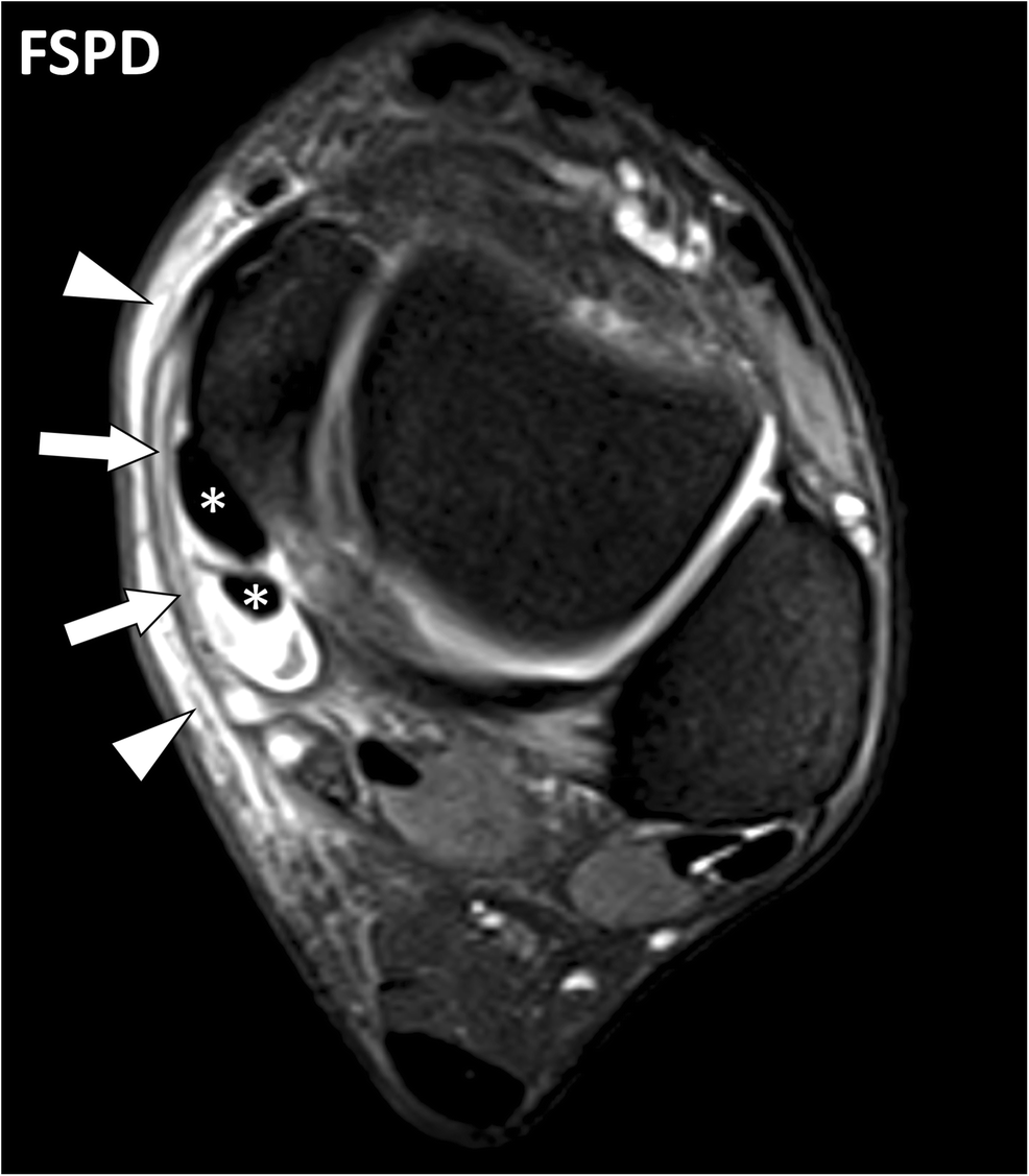
 Fasciae Of The Musculoskeletal System Normal Anatomy And Mr
 Module 2 Lower Extremity Orthopedic Imaging
 Lower Extremity Mri Of Anatomical Atlas
 Radiology Images
 Muscle Mri
 Fasciae Of The Musculoskeletal System Normal Anatomy And Mr


Posting Komentar
Posting Komentar