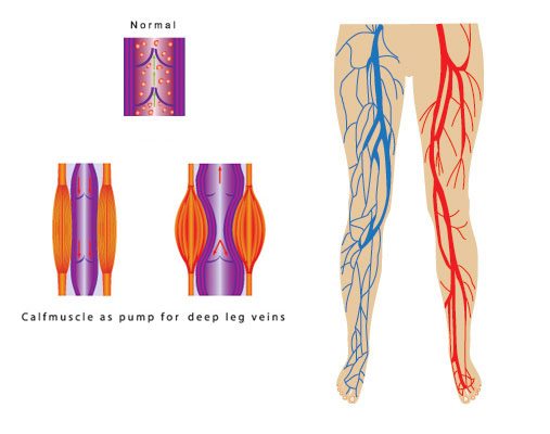
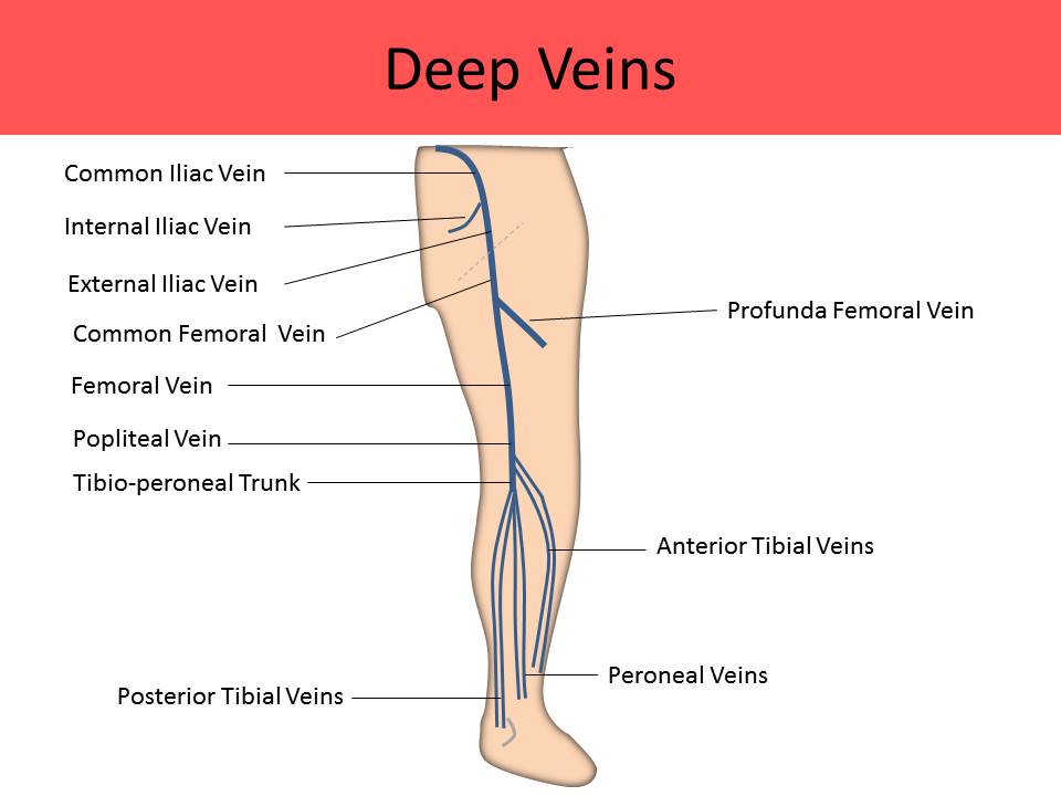
These veins play an important role. Approximately 90 of the venous return in the lower extremities is through the deep veins through the action of the foot calf.
 Anatomy Of Deep Vein Thrombosis
Share your thoughts in the comment section.
Calf veins anatomy. The pressure of blood in the arterial system drops significantly in the feet as energy is lost along the path of flow. Generally the single vein will be slightly larger than if paired. Get a better understanding of the calf vein anatomy with susan gustavson rvt rdms and julie cardoso rdcs rvt rphs.
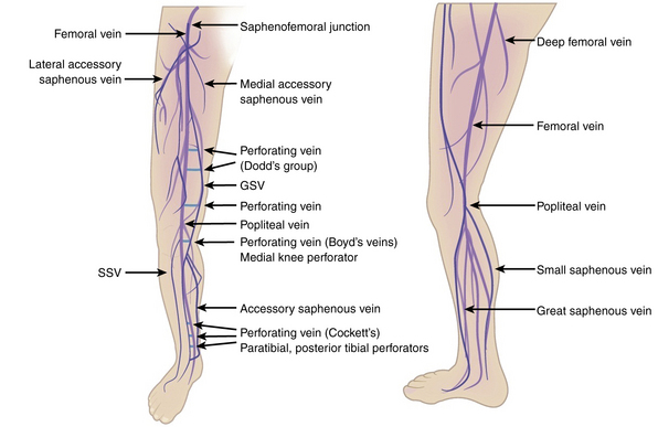
The accumulation of blood in the lower extremity veins while upright is limited by the physical properties of the venous wall the function of the venous valves and the action of the calf muscle pump. The anterior accessory gsv of the leg drains the anterior aspect of the leg below the knee. Learn more with 123sonography.
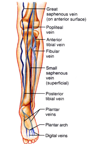
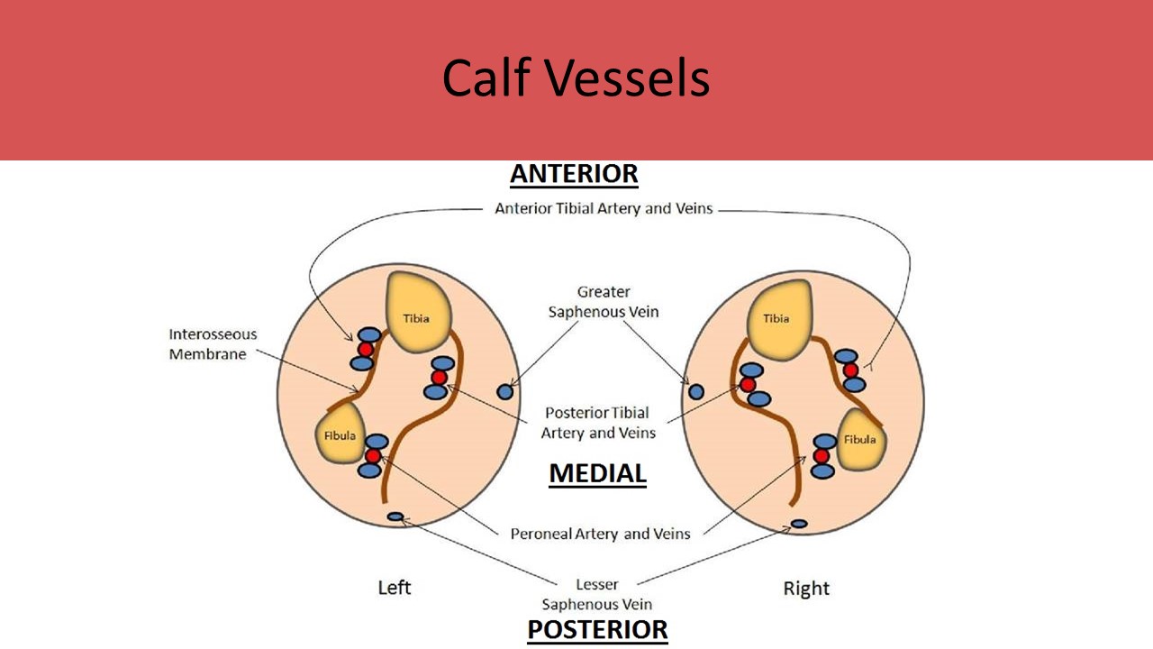
These leg veins have also developed valves to assist in this process. Transversely with the toe of the probe on the medial edge of the mid tibia locate the paired posterior tibial and peroneal veins. Patient still seated as above.
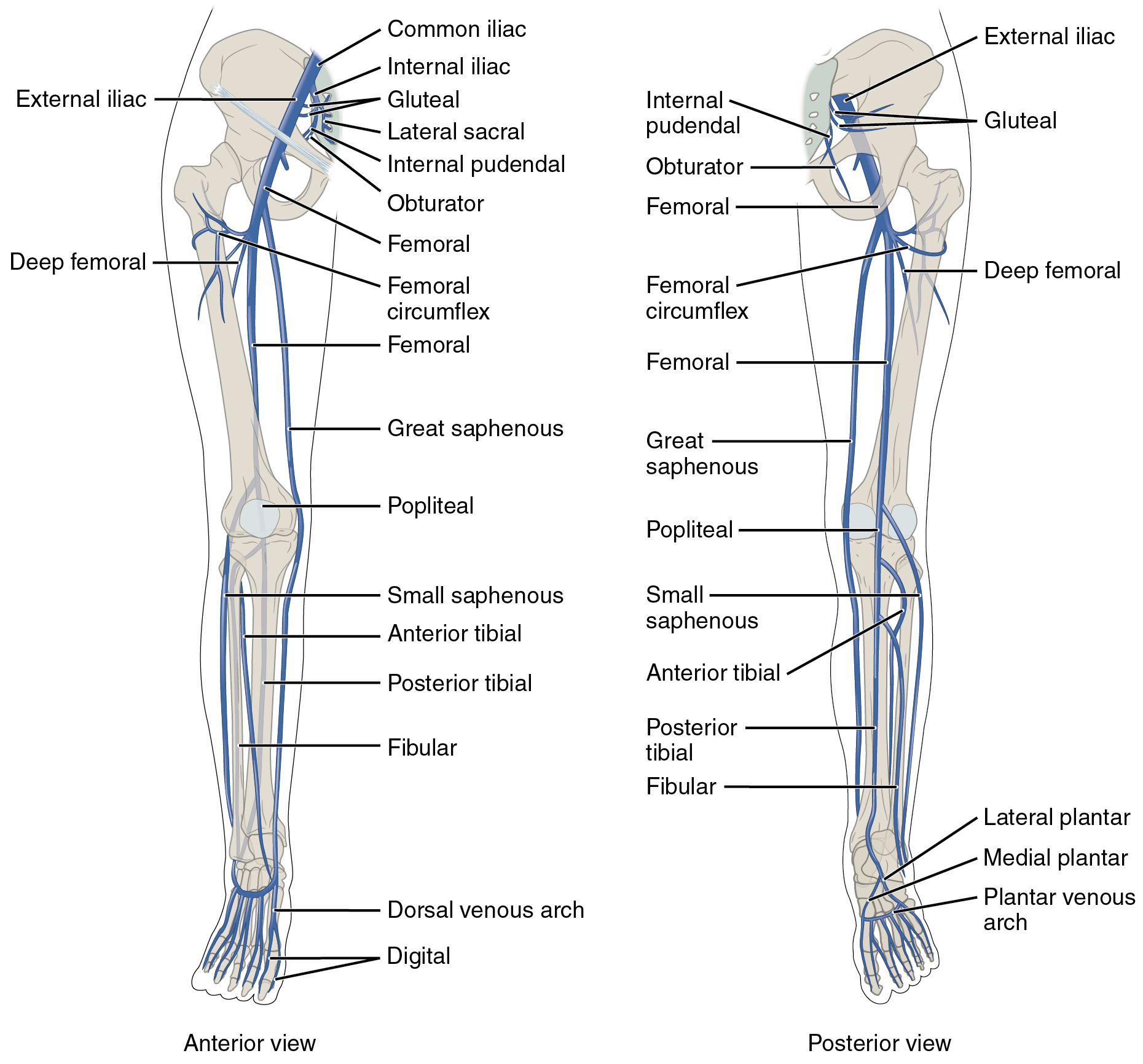
The calf muscle pump. The anterior tibial veins are formed by the venous network of veins on the dorsum of the foot. Venous sinusoids within the calf muscle coalesce to form soleal and gastrocnemius intramuscular venous plexuses which join the peroneal veins in the mid calf.
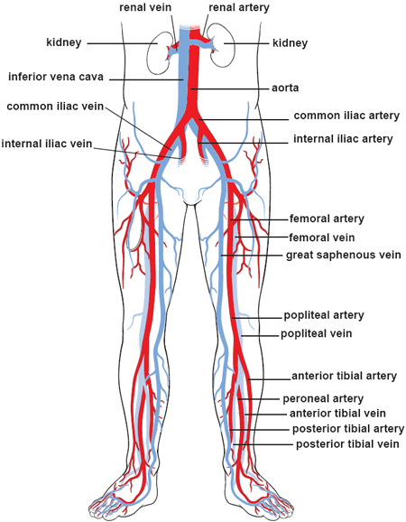
A common variant is for there to be a single vein instead of a pair. Coupled with the counteracting force of gravity the leg veins work along with the leg muscles to return blood back to the heart. They course up the leg between the tibia and the fibula and join the tibialperoneal trunk to form the popliteal vein in the upper calf.
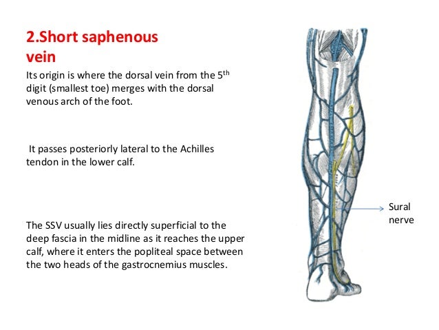
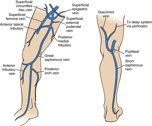
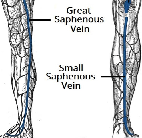
The posterior accessory gsv of the leg leonardos vein or posterior arch vein is a common tributary it begins posterior to the medial malleolus ascends on the posteromedial aspect of the calf and joins the gsv distal to the knee see figure 28. Leg veins thigh lower leg anatomy pictures and names.
 Varicose Vein Can Affect Men Too Great Saphenous Vein

 Leg Veins Canvas Print
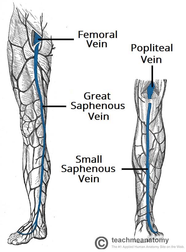 Venous Drainage Of The Lower Limb Teachmeanatomy
 What Is Normal Vein Function
 All About Vein Disease St Louis Varicose Veins And Other
 Ultrasonography
 Varicose Vein Anatomy Pathophysiology Managemant
 Management Of Varicose Veins Thoracic Key
 Deep Vein Thrombosis Wikipedia
 How Leg Veins Work Wisconsin Vein Center Varicose Vein
 Leg Vein Anatomy Veinspecialistsofarizona Com
 Lower Extremity Veins Radiology Key
Is There Any Anastomosis Between Great And Short Saphenous
 Leg Dvt Normal Ultrasoundpaedia
 Varicose Veins Clinical Gate
 Varicose Veins Clinical Features Management
 Anatomy Of The Lower Extremity Veins Varicose Veins
 Diagram Of Gastrocnemius Auto Electrical Wiring Diagram
/vascular-system-veins-56c87fa03df78cfb378b3e7c.jpg) What Is A Vein Definition Types And Illustration
What Is A Vein Definition Types And Illustration
 Ultrasound Registry Review Extremity Venous
 Venous Ulcers Of The Leg Occur Because The Veins Become
Ultrasound Evaluation Of The Lower Extremity Veins
 Ultrasound Registry Review Extremity Venous
 Vascular Lesson 6 Lower Extremity Anatomy Slides 29 48
 Schematic Representation Of Normal Anatomy And Dynamics Of
 Perforator Vein An Overview Sciencedirect Topics
 Drawing Of The Veins Of The Leg And The Calf Muscle Pump
What Are The Peroneal Veins With Pictures
 Vascular Anatomy Of The Calf
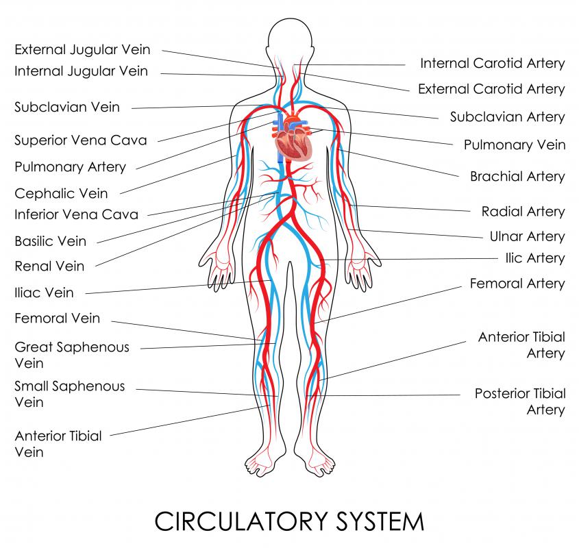
 20 5 Circulatory Pathways Anatomy And Physiology


Posting Komentar
Posting Komentar