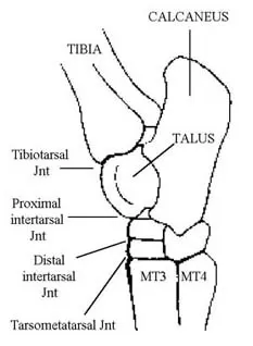
Distal intertarsal joint or centrodistal joint. Although the tarsus refers specifically to the bones and joints of the hock most refer to the hock in such a way as to include the bones joints and soft tissue of the area.
 The Equine Tarsus Hock Vet Physio Phyle
The hock joint allows movement of the hind leg and consists of the tarsus bones the tuber and the calcaneus at the back which forms the point of the hock.
Horse anatomy hock. Cranial the plane going towards the head end front. If he uses his hind end to propel himself and is light on the forehand it will reduce his risk of lameness. Horses and hock problems.
Proximal intertarsal joint or talocalcanealcentroquartal joint. First youll need to know that all this anatomy is based on a median plane. On the other hand a bog spavin is a soft swelling of the front inside corner in the upper half of the hock.
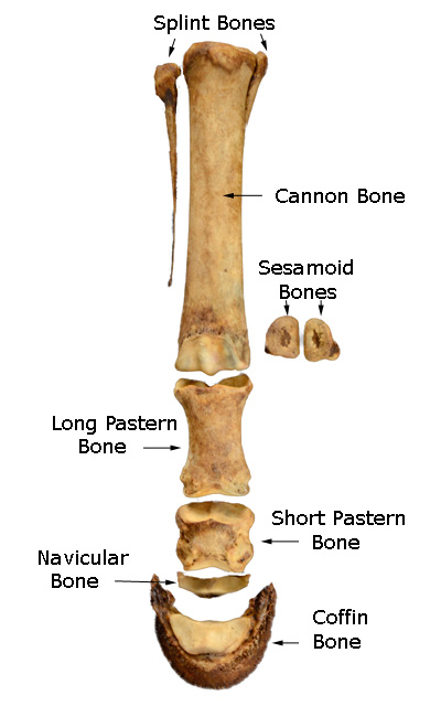
Below the hock joint are the hind cannon with splint bones the long and short pastern the coffin joint and bone the sesamoid bones. The horses hind limbs. This type of spavin is a firm swelling of the inside front corner on the lower half of the hock.
If you continue browsing the site you agree to the use of cookies on this website. The hoof wall is the tough outside covering of the hoof that comes into contact with the ground and is in many respects a much larger and stronger version of the human fingernail. Caudal the plane going towards the hindend.
Horse anatomy muscles of the rear. If he mainly travels on the forehand it can set him up for future lameness. Hock conformation is a very important part of a pre purchase examination.
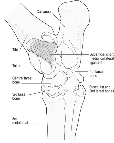
The horses hock is made up of 10 bones and 4 joints supported by several ligaments. Tibiotarsal or tarsocrural joint. Properly conditioned muscles along with good conformation on the hind end will increase the longevity of your horse.
The foot of the horse. Breeders should select for horses with good hock and rear limb conformation. It is similar to the ankle of a human.
The tarsus of the horse hindlimb equivalent to the human ankle and heel the large joint on the hind leg. That means that if you were to draw a line straight down the middle of the back it would divide the horse into equal right and left halves. Tarsus anatomy dane tatarniuk dvm slideshare uses cookies to improve functionality and performance and to provide you with relevant advertising.
Tarsus joint hock the hock is the joint between the tarsal bones and tibia. Joints in the horse. Extra fluid in the joint capsule is the cause of this swelling.
The horses hock joint is one of the hardest working of all the joints and plays a critical role especially in performance horses. It is also one of the most complicated. In the horse the hock consists of multiple joints namely.
As a horse owner you should be familiar with the basic anatomy of the hock and what your horses hocks look like normally.
 Tarsus And Stifle Veterian Key
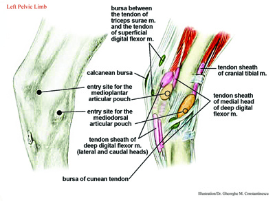
 Novobrace Tendonitis Desmitis And Soft Tissue Injury
 What To Know Before You Inject Instrideedition
 The Horse S Hock Treatments And Symptoms Of Hock Joint
 Radiography Lab Quiz 4 Anatomy And Directional Terms
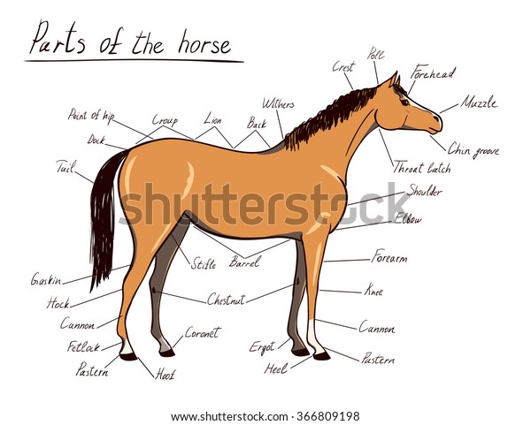
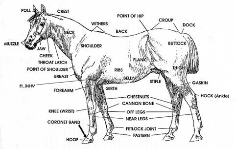
Parts Horse Equine Anatomy Equestrian Scheme Stock Vector
 Horse Anatomy Mobility Health
 Disorders Of The Tarsus In Horses Musculoskeletal System

 Horse Anatomy Mobility Health
How To Interpret Radiographs Of The Carpus And Tarsus Of The
 The Horse S Hock Treatments And Symptoms Of Hock Joint
 Parts Of A Horse Useful Horse Anatomy With Pictures 7 E S L
 On The Injured List Common Problems For Show Horses
 Horse Hock Conformation Hocks Are Important Local Riding
 Tarsal Anatomy Of The Horse
 Horse Anatomy Docx 1 Withers 2 Loin 3 Croup 4 Cannon 5
 Tarsus Aovet Equine Ao Surgery Reference
 Hock Anatomy Wikipedia
 Equine Hock Anterior View
 The Horse Anatomy Workbook A Learning Aid For Students
 Equine Distal Limb Diagnostic Anaesthesia 1 Basic
 Novobrace Tendonitis Desmitis And Soft Tissue Injury
 Distal Tarsitis South Shore Equine Clinic
 Horse Hock Anatomy Horse Anatomy Animal Medicine Horse Care
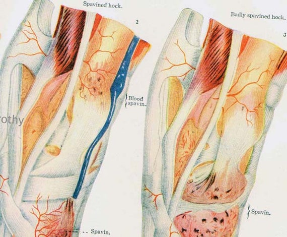 Horse Anatomy Hock Injury Veterinarian Chart 1920s Color Lithograph Antique Large Animal
 Tarsus Equine Anatomy Radiology Small Animal Hospital
 50 Best Equine Anatomy Images Horse Anatomy Anatomy


Posting Komentar
Posting Komentar