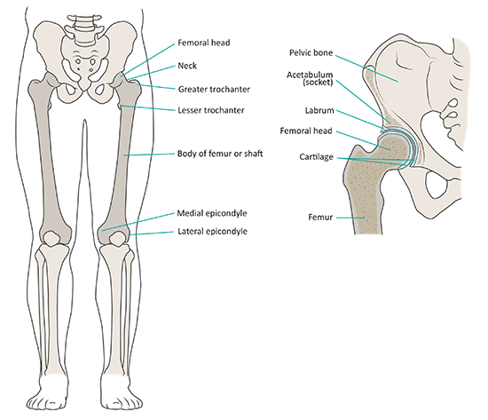
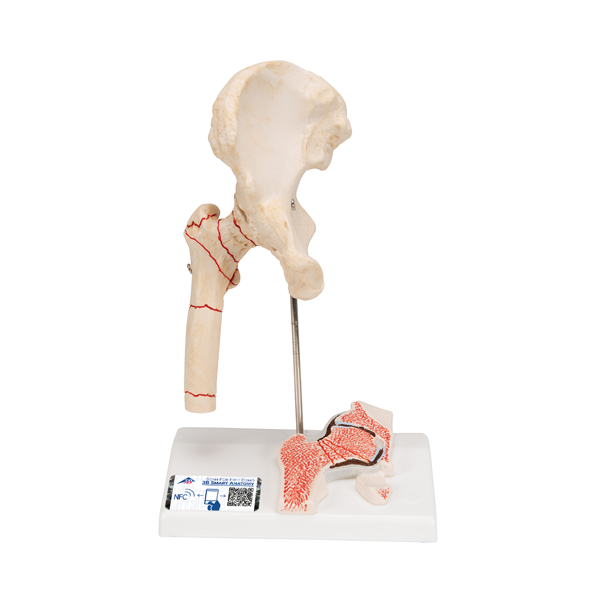
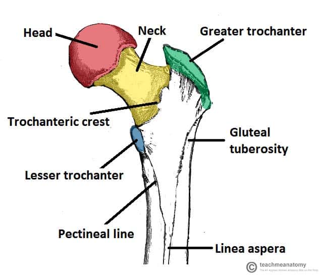
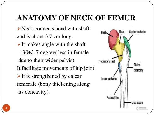
The neck is about is about 3 35 cms long and connects head with the shaft. In the vast majority of cases a hip fracture is a fragility fracture due to a fall or minor trauma in someone with weakened osteoporotic bone.
 Femoral Head Images Stock Photos Vectors Shutterstock
Over 65000 hip fractures each year are recorded in the uk alone and they are becoming increasingly frequent due to an aging population.

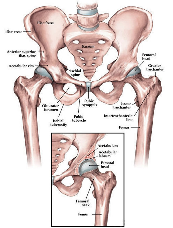
Neck of femur anatomy. A fractured neck of femur nof is a common orthopaedic presentation. Head connects with the acetabulum of the pelvis to make the hip joint. With associated lengthy hospital stays and an estimated cost of over 7500 per fracture.
In the adult the neck forms an angle of about 125 with the body but this varies in inverse proportion to the development of the pelvis and the stature. A fracture of the femoral neck is classified as a type of hip fracture. The angle facilitates movements of the hip joint.
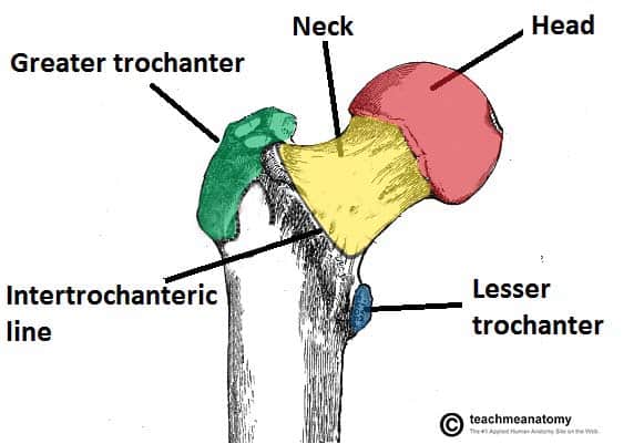
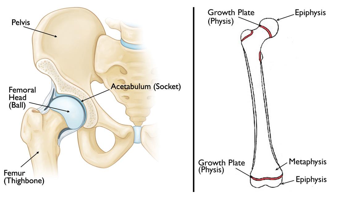
It is often due to osteoporosis. The femoral aspect of the hip is made up of the femoral head with its articular cartilage and the femoral neck which connects the head to the shaft in the region of the lesser and greater. The proximal end consists of a head neck and two trochantersthe head faces superiorward medialward and slightly anteriorward the proximal area of the femur forms the hip joint with the pelvis.
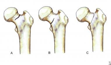
The neck forms an angle with the shaft known as neck shaft angle and is about 125 in adults lesser in females. The classical clinical finding is that of an externally rotated shortened leg. Fractures of the neck of femur are very common injuries which mainly occur in elderly females with osteoporotic bones.
There are also two bony ridges connecting the two trochanters. In the female in consequence of the increased width of the pelvis the neck of the femur forms more nearly a right angle with the body than it does in the male. A fractured neck of femur is classified as either intracapsular or extracapsular.
The femoral neck is strengthened by a thickening of bone called the calcar femorale present along its concavity. The head forms a ball and socket joint with the hip at the acetabulum being held in place by a ligament ligamentum teres femoris within the socket and by strong surrounding ligaments. In humans the neck of the femur connects the shaft and head at a femur upper bone of the leg or hind leg.
 The Femur Proximal Distal Shaft Teachmeanatomy
 Femur Anatomy Britannica
 Neck Of Femur Fracture Subcapital Intertrochanteric
 Royalty Free Femur Stock Images Photos Vectors Shutterstock
 Femoral Osteotomy An Overview
 Management Of Acute Hip Fracture Nejm
 Slipped Capital Femoral Epiphysis Orthoinfo Aaos
 Anatomical Teaching Models Plastic Human Joint Models
 Human Femur Anatomy With Porosity And Stiffness At Different
 Hip Fracture Anatomy Causes And Consequences Intechopen
 Femoral Nerve Block Landmarks And Nerve Stimulator
 Femoral Neck Fracture Background Epidemiology Functional
 Transcervical Femoral Neck Fractures Causes Treatment Rx
 The Femur Proximal Distal Shaft Teachmeanatomy
 The Femur Human Anatomy
Transient Osteoporosis Of The Hip Orthoinfo Aaos
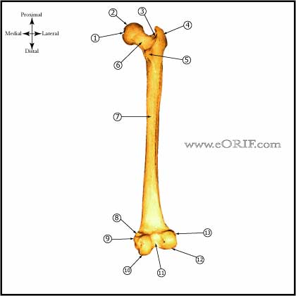 Femur Anatomy Eorif
 Femoral Neck Fracture Hip Fracture Physioadvisor
 Anatomy Of The Hip Central Coast Orthopedic Medical Group
 Fracture Neck Of Femur
![]() Femur Bone Anatomy Proximal Distal And Shaft Kenhub
Femur Bone Anatomy Proximal Distal And Shaft Kenhub
 Hip Canadian Orthopaedic Foundation Canadian Orthopaedic
 Biomechanics Of Femoral Neck Fractures In Runners Lower
 Dr Tooth Care Anatomy Of Femur Bone
 Startradiology
 Femoral Neck Stress Fractures Knee Sports Orthobullets

 Femoral Head Fracture
 Fracture Neck Of Femur


Posting Komentar
Posting Komentar