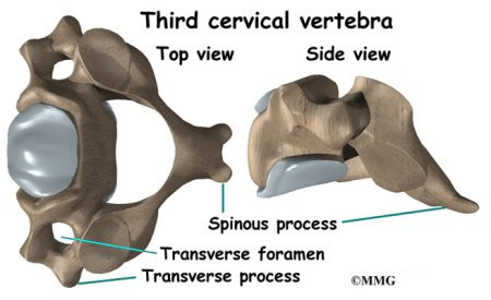
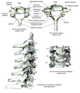
The c1 vertebra is formed like a ring that sits on top of c2. There are seven cervical vertebrae in the human body.
 Anatomical Teaching Models Plastic Vertebrae Model
The dens forms a joint with the c1 vertebra and facilitates its turning motions thereby allowing the head to turn in different directions.
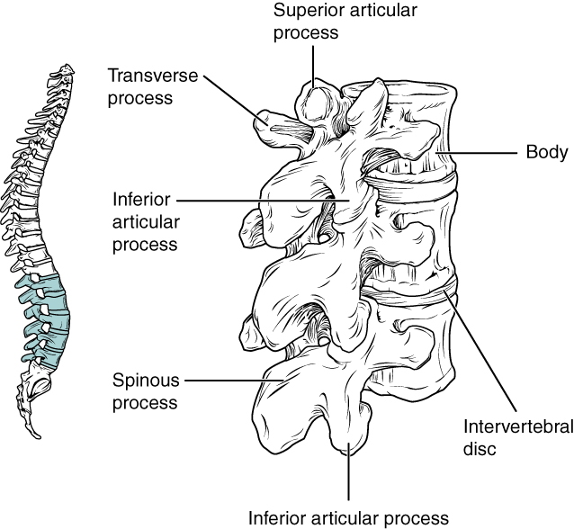
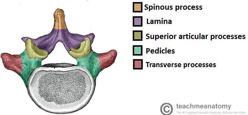
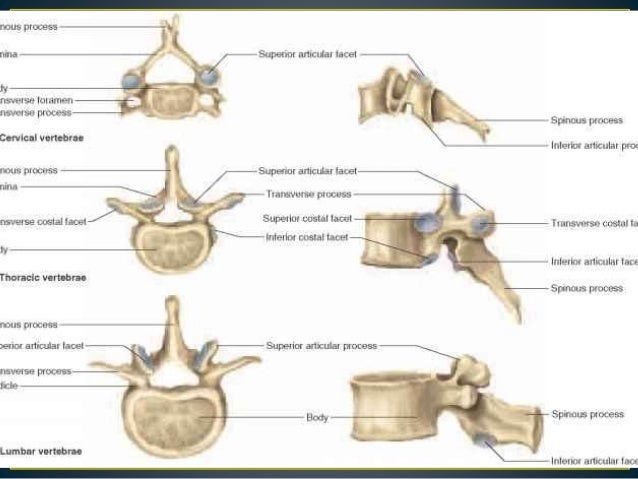
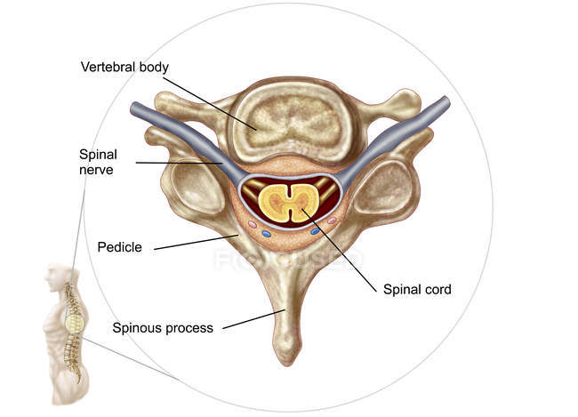
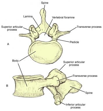
Anatomy of a vertebra. Support of the vertebrae function in the skeletomuscular system by forming. They also protect the delicate spinal cord and nerves within their vertebral canal. Vertebrae contain a vertebral foramen for the passage of the spinal canal.
Structure of a vertebrae. This bony knob is called the odontoid process or dens. The vertebral arch is posterior meaning it faces the back of a person.
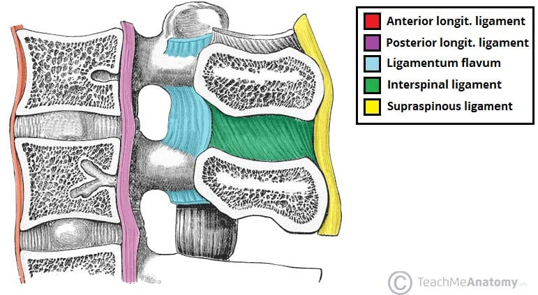
The c2 vertebra has an upward facing long bony process called the dens. All vertebrae share a basic common structure. Muscles ligaments and discs attach to various parts of a vertebra.
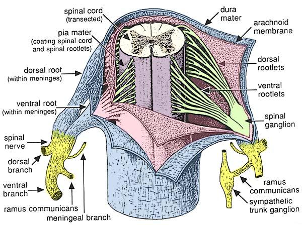
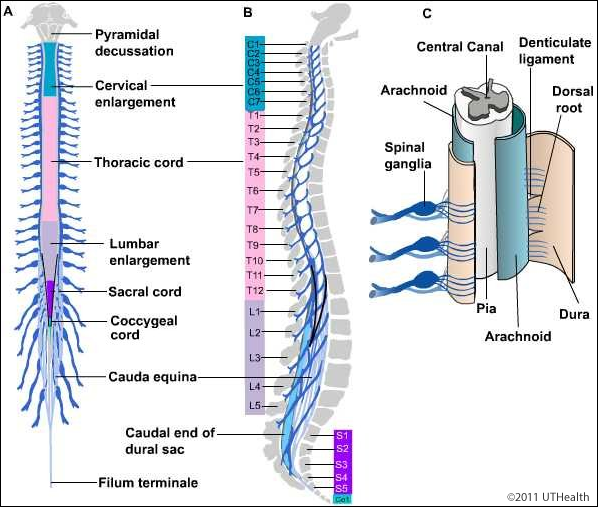
The space between the vertebral body and the arch is the spinal canal for your spinal cord. The joint between the c1 and c2 vertebrae is called the atlantoaxial joint. The vertebral column functions.
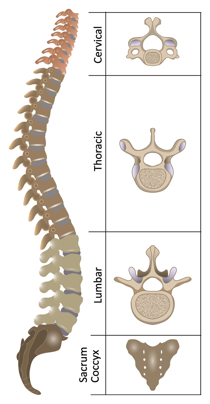
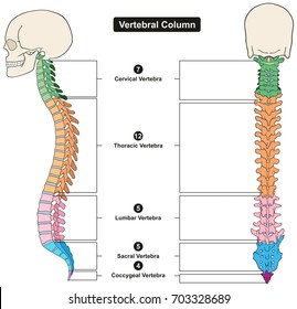
Seen from above a vertebra looks like a giant head with three pieces sticking out and a hole in the middle. Cervical thoracic lumbar sacrum and coccyx fig. The vertebrae also provide the openings the intervertebral foramina which allow.
The vertebrae are numbered and divided into regions. Together these enclose the vertebral foramen which contains the spinal cord. Anatomy of a vertebra a typical vertebra consists of two parts.
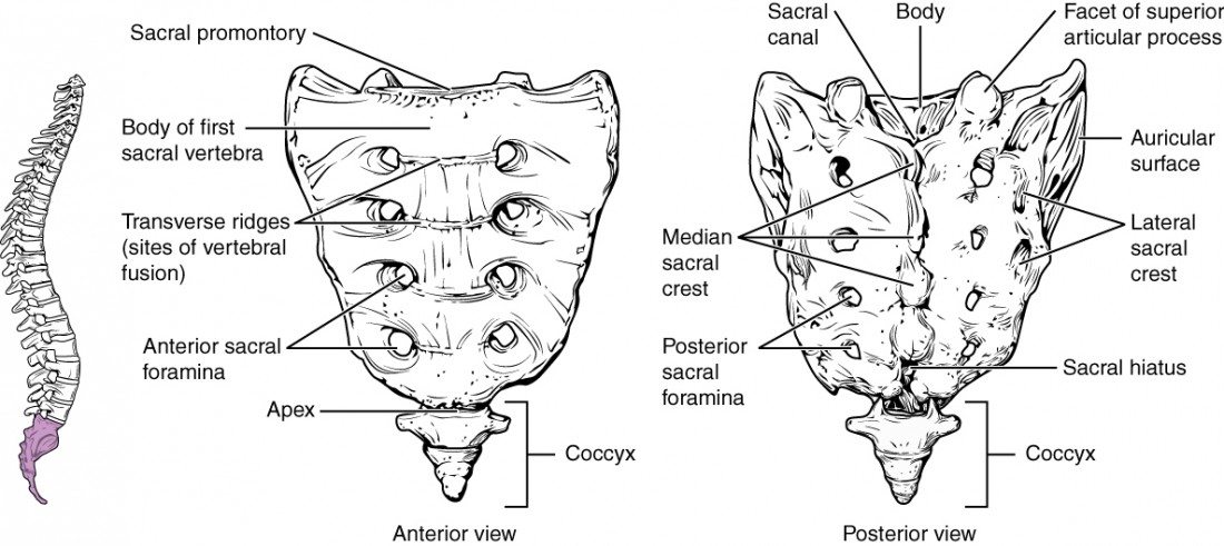
Functions of vertebrae include. The vertebrae of the sacrum and coccyx are fused. Vertebrae are the 33 individual bones that interlock with each other to form the spinal column.
The vertebral body and the vertebral arch. These two vertebrae have different anatomy than the rest of the spine. The c2 vertebra has a bony knob that fits into the front portion of the ring of the c1 vertebra.
Protection encloses and protects the spinal cord within the spinal canal. Only the top 24 bones are moveable. These vertebrae carry all of the upper bodys weight while providing flexibility and movement to the trunk region.
The lumbar vertebrae consist of five individual cylindrical bones that form the spine in the lower back.
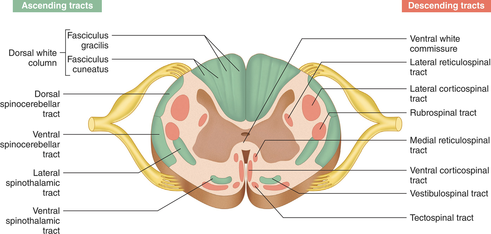 Functional Anatomy Of The Spinal Cord Springerlink
 The Vertebral Column Joints Vertebrae Vertebral Structure
 The Vertebral Column Anatomical Poster Size 24wx36t Amazon
 Anatomy Of A Human Vertebra The Image Is Adopted From 6

![]() Spinal Cord Anatomy Structure Tracts And Function Kenhub
Spinal Cord Anatomy Structure Tracts And Function Kenhub
 Spinal Cord Anatomy Spine Orthobullets
 Anatomy For First Aiders The Spinal Cord First Aid For Free
 The Vertebral Column Joints Vertebrae Vertebral Structure
 7 3 The Vertebral Column Anatomy And Physiology
 The Vertebral Column Anatomy And Physiology I
 Spinal Anatomy Vertebral Column
 Anatomy Of Spinal Stenosis
 The Vertebral Column Anatomy And Physiology I
 Skull Anatomy Vertebral Column Cervical Vertebrae Central
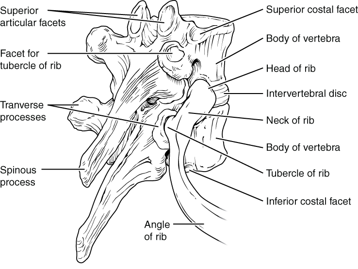
 Anatomy Of Thoracic Vertebra
7 3 The Vertebral Column Anatomy And Physiology
 Spine Anatomy Goodman Campbell Brain And Spine
 Anatomy Of The Brain And Spinal Cord Seattle Cancer Care
 Medical Illustration Of Human Vertebra Anatomy Spinal Cord
 Cervical Spine Anatomy Eorthopod Com
 Anatomy Of The Spine Spinal Cord Injury Information Pages
 Neuraxial Anatomy Nysora
 Cervical Spine Anatomy Overview Gross Anatomy
 Lumbar Spine Anatomy Overview Gross Anatomy Natural Variants
 Gross Anatomy Vertebral Column Flashcards Quizlet
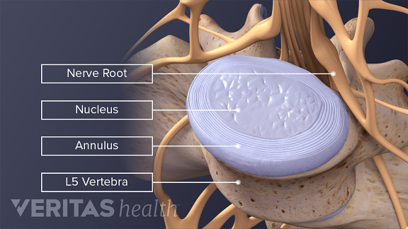 Spinal Discs
![]() Vertebral Column Anatomy Vertebrae Joints Ligaments
Vertebral Column Anatomy Vertebrae Joints Ligaments
 Notes On Anatomy And Physiology The Vertebrae
 Neuroanatomy Online Lab 4 External And Internal Anatomy
 Vertebral Column Images Stock Photos Vectors Shutterstock
 Spinal Cord Anatomy Healthlink Bc
 Spinal Anatomy Vertebral Column


Posting Komentar
Posting Komentar