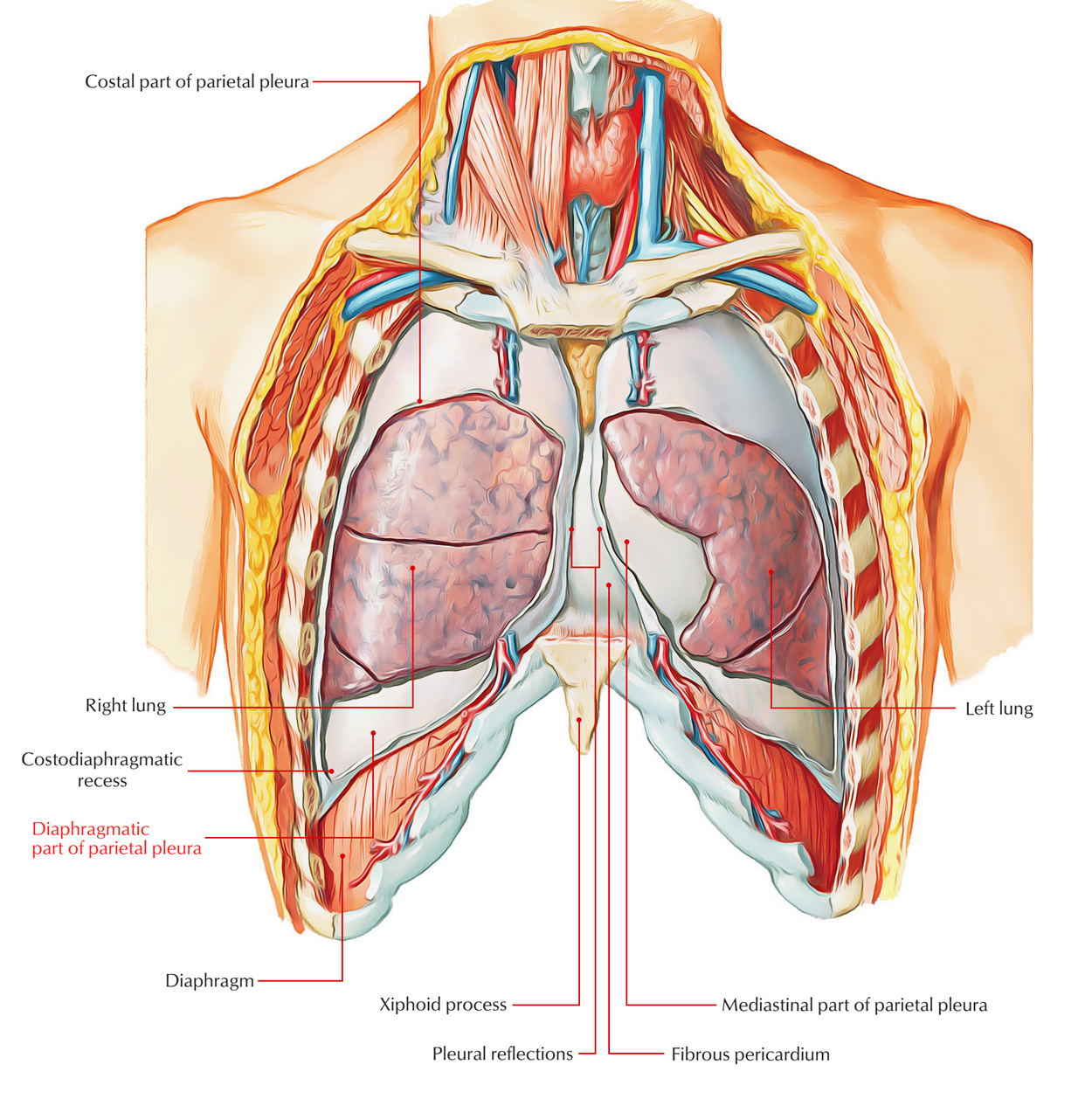
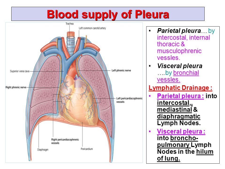
Each lung is invested by an exceedingly delicate serous membrane the pleura. Parietal pleura covers the internal surface of the thoracic cavity.
 Normal Anatomy Of Pleural Cavity And Lungs Medical Art Works
Parietal pleura the portion of the pleura external to the pulmonary pleura lines.

Anatomy of pleura. The parietal pleura folds back on itself at the root of the lung to become the visceral pleura. For the best protection and function of the lung. The pleural cavity also known as the pleural space is the thin fluid filled space between the two pulmonary pleurae known as visceral and parietal of each lung.
Sharmin susiwala ty bpt cts. Page 2 borders of the lungborders of the lung. The apexthe apex is about 2 3 cms 1 inch.
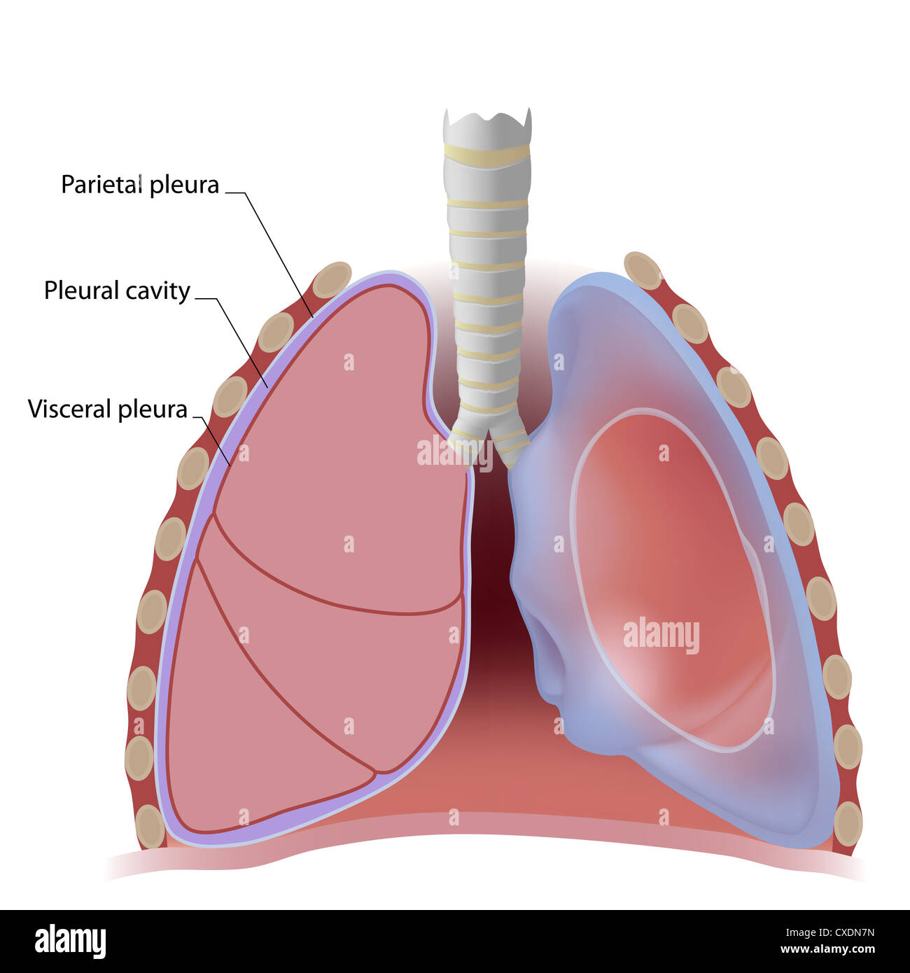
The pleura is a serous membrane which folds back onto itself to form a two layered membrane structure. The outer pleura is attached to the chest wall 1 9. It comprises of two layers outer parietal and inner visceral layer.
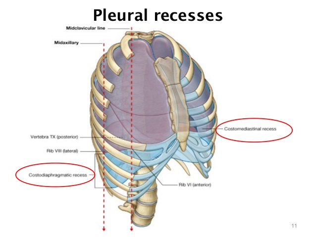
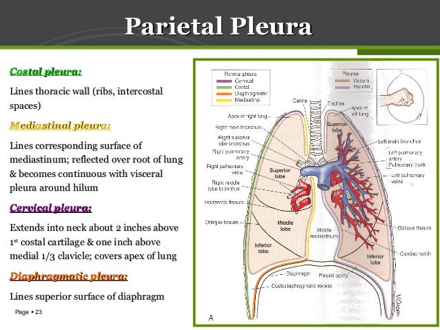
It normally contains only a thin film of serous fluid which is secreted by the pleura. The thin space is known as the pleural cavity and contains a small amount of pleural fluid few milliliters in a normal human. Each pleura can be divided into two parts.
Anatomy of the pleura. Within the confined spaces vital organs such as the heart and lung however have to move and change volume continuously to function. The lower.
A small amount of fluid between these layers roughly 4 to 5 cc of pleural fluid helps to act as a cushion. Function of the pleura. In health the two pleurae are in contact.
Page 3 borders of the lungborders of the lung. The pleura is not the only set of membranes lining body cavities. Pleural cavity is a closed potential space between the parietal and visceral layers of pleura.
Visceral pleura covers the lungs. Anatomy of pleura grays description simplified 1. Pleural cavity the pleural cavity is the space lined by a serous membrane called the pleural membrane the membrane covers both the lungs and the thoracic wall.
A pleura is a serous membrane which folds back onto itself to form a two layered membranous pleural sac. In humans all vital organs are protected within a body wall formed by ribs vertebrae and layers of thick muscle. Pleura is a serous membrane lined by mesotheliumsimple squamous epithelium that is present as a closed sac around the lungs.
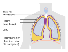
The potential space between the two membranes is the pleural cavity it contains a thin layer of fluid which helps in. When the lung collapses however or when air or liquid collects between the two membranes the pleural cavity or sac becomes apparent see pleurisy. Iman galal mdiman galal md pulmonary medicine departmentpulmonary medicine department ain shams.
Anatomy of lung pleura 1. There is a membrane around the heart pericardium and a membrane lining the abdominal cavity peritoneum as well.
 Pleural Cavities Anatomy Of The Respiratory System
 Diagnosis And Management Of Patients With Pleural Effusions
 Diaphragmatic Pleura Earth S Lab

 Drawing To Show The Anatomy Of Normal Healthy Lung And Pleurae
 Overview Of Benign Pleural Conditions Anatomy And
 Anatomy Of Pleura Grays Description Simplified
 Lung Pleura And Pleural Cavity Stock Photo 50703849 Alamy
Surface Anatomy Of The Pleura And Thoracocentesis
 Anatomy Of Pleura Grays Description Simplified
 Anatomy Of Pleura Grays Description Simplified

 Anatomy Of Lung Pleura
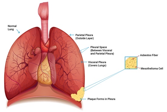 Pleural Mesothelioma Symptoms Prognosis And Top Treatments
 Pleural Space Part 1 Of 3 Hd
 Pdf Pleura Space Anatomy Semantic Scholar
 Pleura And Lung By Prof Saeed Abuel Makarem Dr Sanaa Al
 Figure Pleura Visceral Pleura Left Lung Statpearls
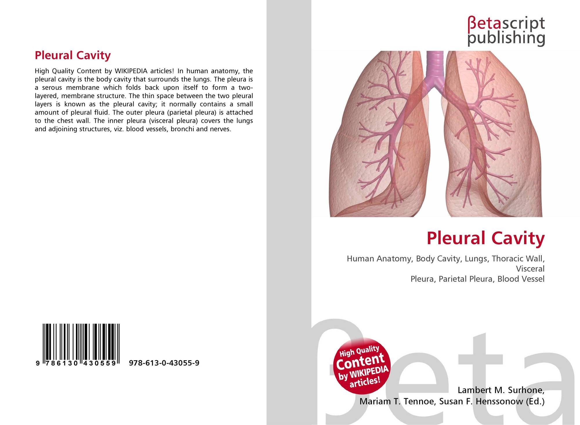 Pleural Cavity 978 613 0 43055 9 6130430558 9786130430559
 Pleura Of The Lungs Healthlink Bc
 Pleural Cavity Pleura Acland S Video Atlas Of Human Anatomy

 Pleural Cavity Wikipedia
How Do We Breathe Lungs And Pleura Interactive Biology

 Pleural Cavity Wikipedia
 Visceral Pleura Planes Sections And Cavities Cavities


Posting Komentar
Posting Komentar