The anatomy of the posterior aspect of the knee biomechanical analysis of an isolated fibular lateral collateral ligament reconstruction using an autogenous semitendinosus graft effect of tibial positioning on the diagnosis of posterolateral rotatory instability in the posterior cruciate ligament deficient knee. The knee joins the thigh bone femur to the shin bone tibia.
 Posterior Knee Anatomy Gray S Anatomy Illustration
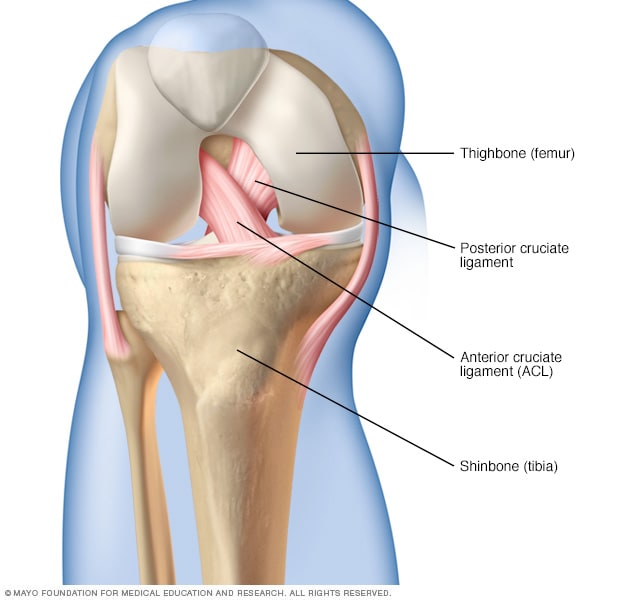
The important ligaments of the knee are the acl pcl lcl and mcl.
Anatomy posterior knee. Knee pain is more common in the anterior medial and lateral aspect of the knee than in the posterior aspect of the knee. Injuries to the posterolateral corner can be debilitating to the person and require recognition and treatment to avoid long term consequences. Most commonly the anterior cruciate ligament and posterior cruciate ligament.
If used to describe the patella knee cap then it would refer to the side of the patella closest to the femur. Injuries to the plc often occur in combination with other ligamentous injuries to the knee. A lack of familiarity leads to hesitancy when performing approaches in these areas of the knee.
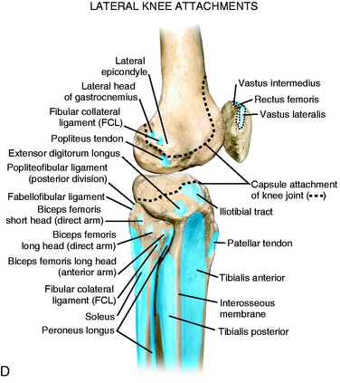
Knowledge of the bony topography will result in a greater number of anatomic ligament reconstructions. The anteroposterior ap position of the itb with knee flexion contributes to the pivot shift phenomena with an anterior cruciate ligament acl. Sciatic pain which radiates down into the back of your leg knee andor lower leg.
Posterior knee pain can be caused by injuries or dysfunction in the lower back and hips. Posterolateral corner injuries of the knee are injuries to a complex area formed by the interaction of multiple structures. Posterior knee pain is a common patient complaint.
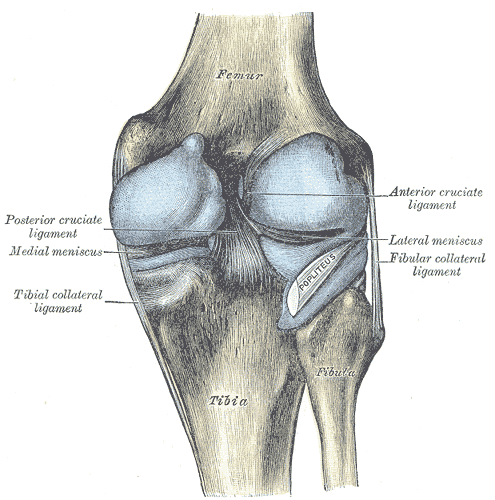
We are pleased to provide you with the picture named posterior view of knee joint. As with any injury an understanding of the anatomy and functio. The acl connects the medial border of the lateral femoral condyle to the anterior aspect of the tibia while the pcl connects the lateral border of the medial femoral condyle to the posterior aspect of the tibia.
Medial the side of the knee that is closest to the other knee if you put your knees together the medial sides of each knee would touch. You will also find anterior cruciate ligament lateral epicondyle posterior meniscofemoral ligament lateral meniscus fibular collateral ligament posterior ligaments head of the fibula as well. The knee is one of the largest and most complex joints in the body.
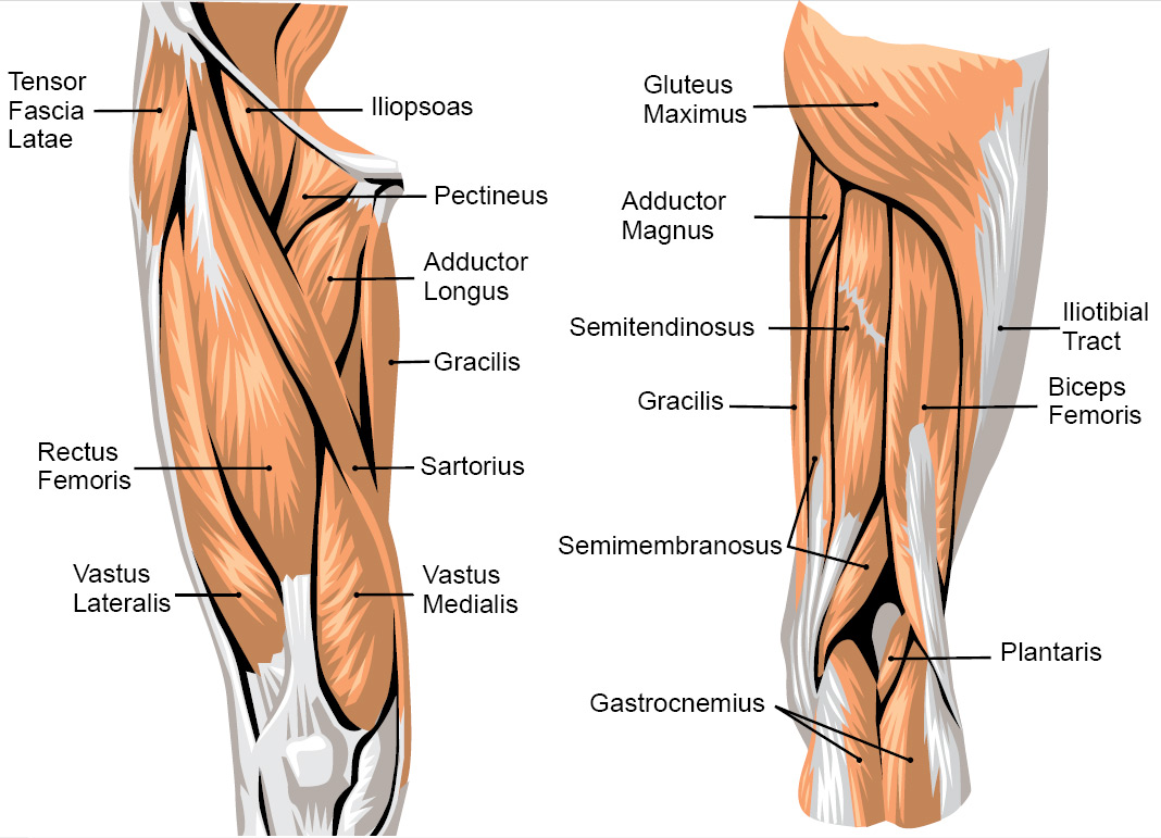
The differential diagnoses for posterior knee pain include pathology to the bones musculotendinous structures ligaments andor to the bursas. When the knee is flexed to 90 the itb moves posterior to the axis of rotation. The slump test is to identity sciatic type referred pain referred pain.
In knee extension the itb is anterior to the axis of rotation and helps maintain extension. Posterior if facing the knee this is the back of the knee. The posterior and lateral anatomy of the knee joint presents a challenge to even the most experienced knee surgeon.
 Posterior Cruciate Ligament Pcl Injury Symptoms And
 Knee Posterior Approach Approaches Orthobullets
 Gastrocnemius Muscle Anatomy Britannica
 Posterior Cruciate Ligament An Overview Sciencedirect Topics
 What Is A Hamstring Strain And What Are The Causes Of A
 Knee Posterior Approach Approaches Orthobullets
 Free Art Print Of Posterior View Of The Right Knee
 Keeping On Track With Knees Expanding Light
:watermark(/images/watermark_only.png,0,0,0):watermark(/images/logo_url.png,-10,-10,0):format(jpeg)/images/anatomy_term/arteria-superior-lateralis-genus/DKWU3EDjKXvcLhiC10ClLg_ToqtbQrWTA_Arteria_superior_lateralis_genus_1.png) Popliteal Artery Anatomy Branches Location And Course
Popliteal Artery Anatomy Branches Location And Course
 Popliteal Ligament An Overview Sciencedirect Topics
 Illitibial Band Syndrome Symptoms Treatment Exercises
 Muscles Advanced Anatomy 2nd Ed
 Inner Knee Pain Why Does The Inside Of My Knee Hurt
 Ultrasound Guided Popliteal Sciatic Block Nysora
Posterior Cruciate Ligament Injuries Orthoinfo Aaos
 The Knee Resource Posterolateral Corner Injury
 Posterior Knee Pain Physiopedia
 Anatomy Of The Knee Bones Muscles Arteries Veins Nerves
 Lateral Posterior And Cruciate Knee Anatomy Clinical Gate
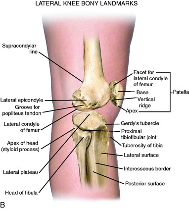 Lateral Posterior And Cruciate Knee Anatomy Clinical Gate
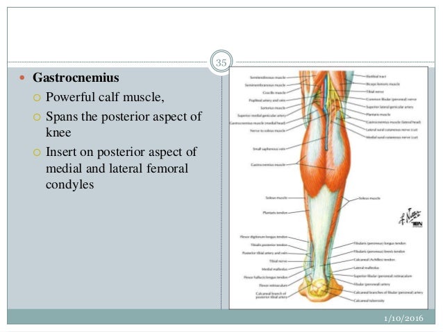 Surgical Anatomy Of Knee Joint
 Muscles Of The Knee Anatomy Pictures And Information
:background_color(FFFFFF):format(jpeg)/images/library/11153/muscles-tibia-fibula_english__2_.jpg) Leg And Knee Anatomy Bones Muscles Soft Tissues Kenhub
Leg And Knee Anatomy Bones Muscles Soft Tissues Kenhub
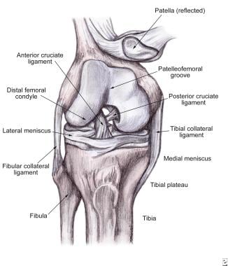 Soft Tissue Knee Injury Practice Essentials Background
 Posterolateral Corner Injury Knee Sports Orthobullets
 Knee Human Anatomy Function Parts Conditions Treatments
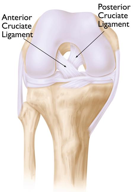 Knee Replacement Implants Orthoinfo Aaos
 Vector Illustration Posterior View Of The Right Knee Eps
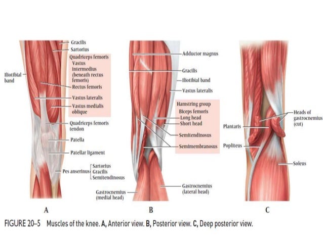 Dr Anurag Applied Anatomy Of Knee


Posting Komentar
Posting Komentar