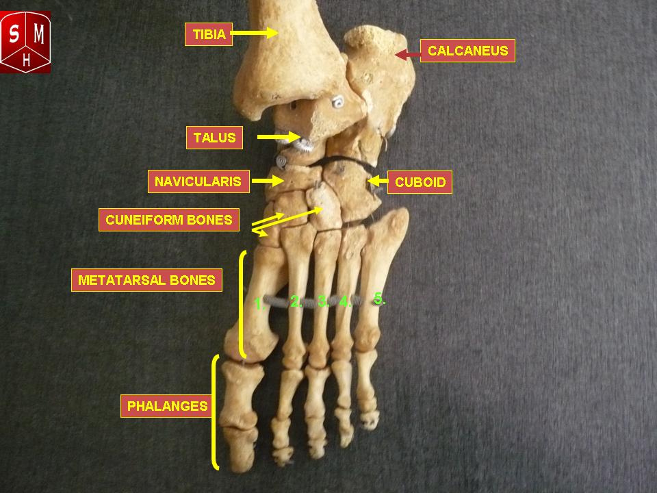
This may sound like overkill for a flat structure that supports your weight but you may not realize how much work your foot does. The cuboid is on the lateral side opposite the arch of the foot and in front of the calcaneus.

The foot consists of thirty three bones twenty six joints and over a hundred muscles ligaments and tendons.
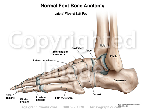
Left foot anatomy. The calcaneus third cuneiform and fourth and fifth metatarsals. The foot contains 26 bones 33 joints and over 100 tendons muscles and ligaments. Pain swelling redness and bruising may be signs of a fracture.
Talus the bone on top of the foot that forms a joint with the two bones of the lower leg the tibia and fibula. Occasionally with a fifth the navicular. The foot is responsible for balancing the bodys weight on two legs a feat which modern roboticists are still trying to replicate.
The structure of the foot is similar to that. Foot and ankle anatomy is quite complex. These all work together to bear weight allow movement and provide a stable base for us to stand and move on.
The navicular sits at the medial side of the tarsus. The cuboid forms a joint with four bones. The foots complex structure contains more than 100 tendons ligaments and muscles that move nearly three dozen joints while bones provide structure.
The foot is an extremely complex anatomic structure made up of 26 bones and 33 joints that must work together with 19 muscles and 107 ligaments to execute highly precise movements. A viral infection in the sole of the foot that can form a callus with a central dark spot. The bones of the feet are.
Tarsals five irregularly shaped bones of the midfoot that form the foots arch. Calcaneus the largest bone of the foot which lies beneath the talus to form the heel bone. At the same time the foot must be strong to support more than 100000 pounds of pressure for every mile walked.
The metatarsal bones are the most frequently broken bones in the feet either from injury or repetitive use.
 Left Foot Anatomical Jewelry Pendant Anatomy Medicine
 Progression Of Left Foot Injury Medical Illustration
 13245 01x The Sesamoid Bones Of Left Foot Anatomy Exhibits
 Muscles Of The Leg And Foot Classic Human Anatomy In
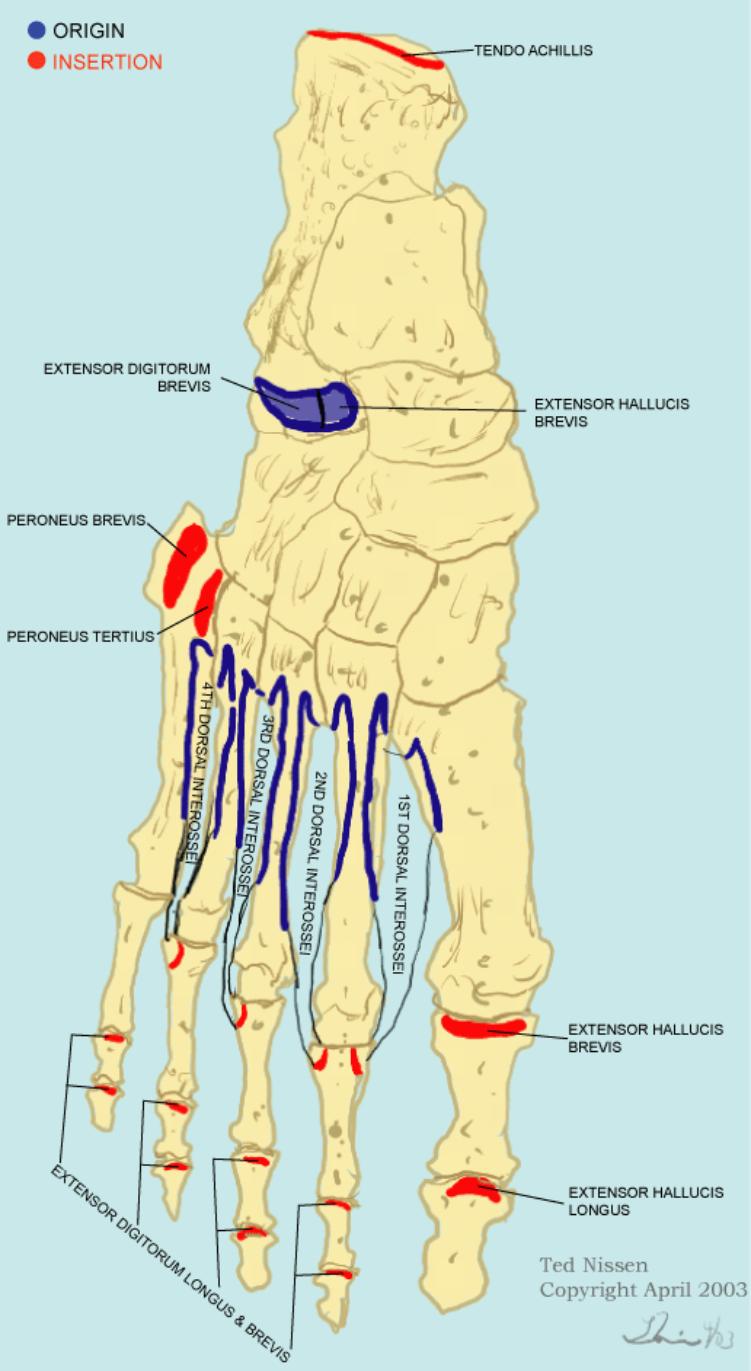
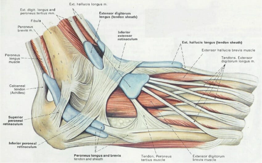
 195 Dorsum Of Left Foot With Muscles And Tendon Sheaths
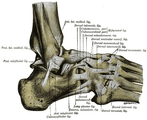
Lateral View Of Left Ankle
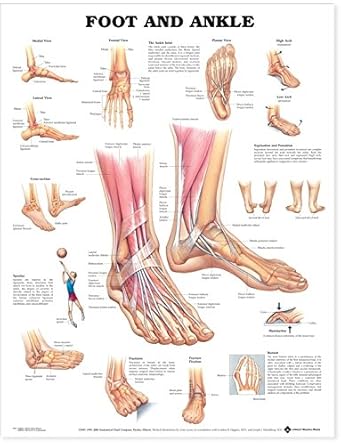 Foot And Ankle Anatomical Chart
 Foot Anatomy Bones Ligaments Muscles Tendons Arches
 Foot Anatomy Bones Ligaments Muscles Tendons Arches
 Physical Therapy In Columbia For Foot Anatomy
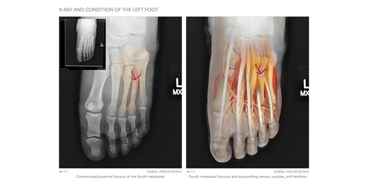 Left Foot X Ray High Impact Visual Litigation Strategies

Anatomical Landmarks On The Dorsum Of The Left Foot Showing
 Human Foot Anatomy Left Leg Inside View Bones Of The Foot
 Library Trial Exhibits Inc


 Left Foot Normal Anatomy Medial View Medical Art Works
 Cross Sections Of The Left Foot On Behance

 Foot Anatomy Bones Ligaments Muscles Tendons Arches
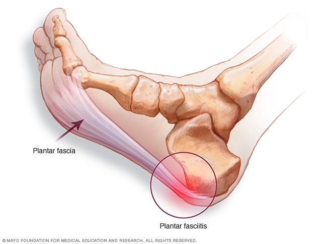 Plantar Fasciitis Symptoms And Causes Mayo Clinic
 3b Scientific A31 1l Human Left Loose Foot And Ankle Skeleton
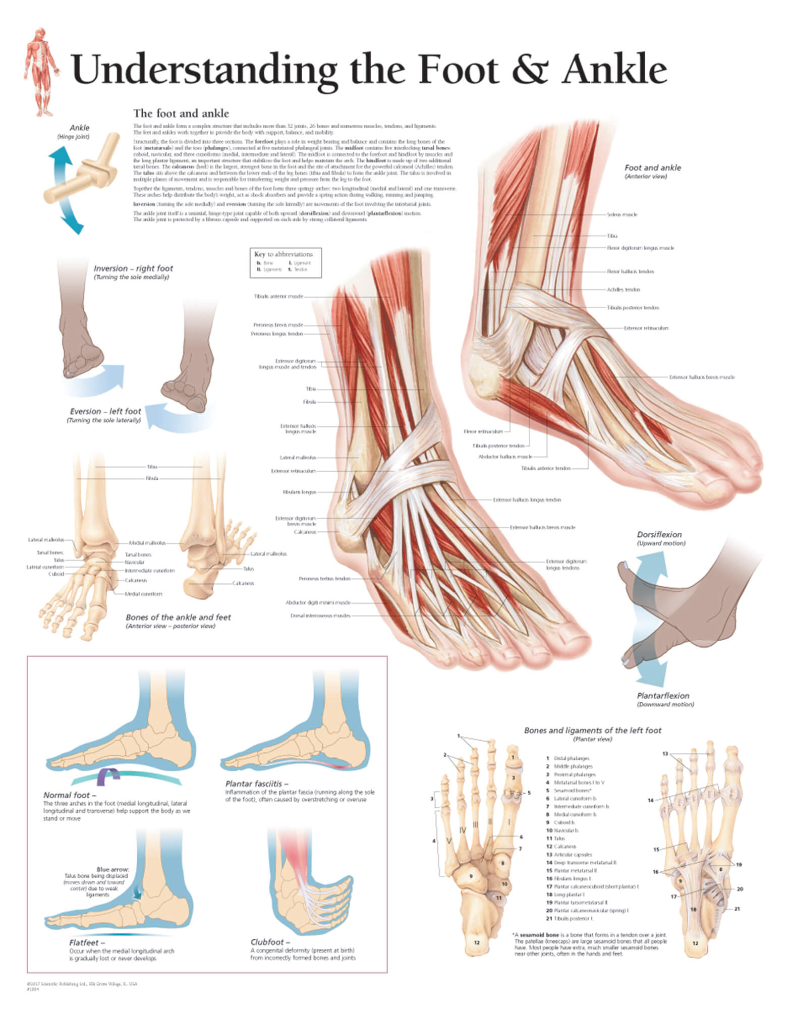 Understanding The Foot Ankle
%2C445%2C291%2C400%2C400%2Carial%2C12%2C4%2C0%2C0%2C5_SCLZZZZZZZ_.jpg) Anatomy And Injuries Of The Foot And Ankle 9781587798375
Anatomy And Injuries Of The Foot And Ankle 9781587798375


Posting Komentar
Posting Komentar