Problems with nerves in the feet are very common. It connects the spinal cord with the outside of the thigh the hamstring muscles in the back of the thighs and muscles in the lower leg and feet.
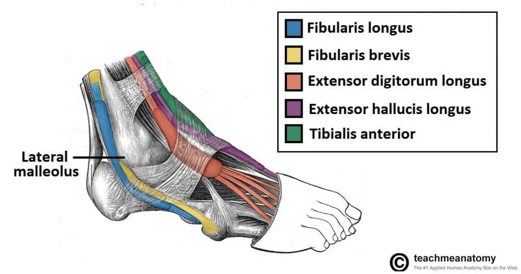 Muscles In The Lateral Compartment Of The Leg Teachmeanatomy
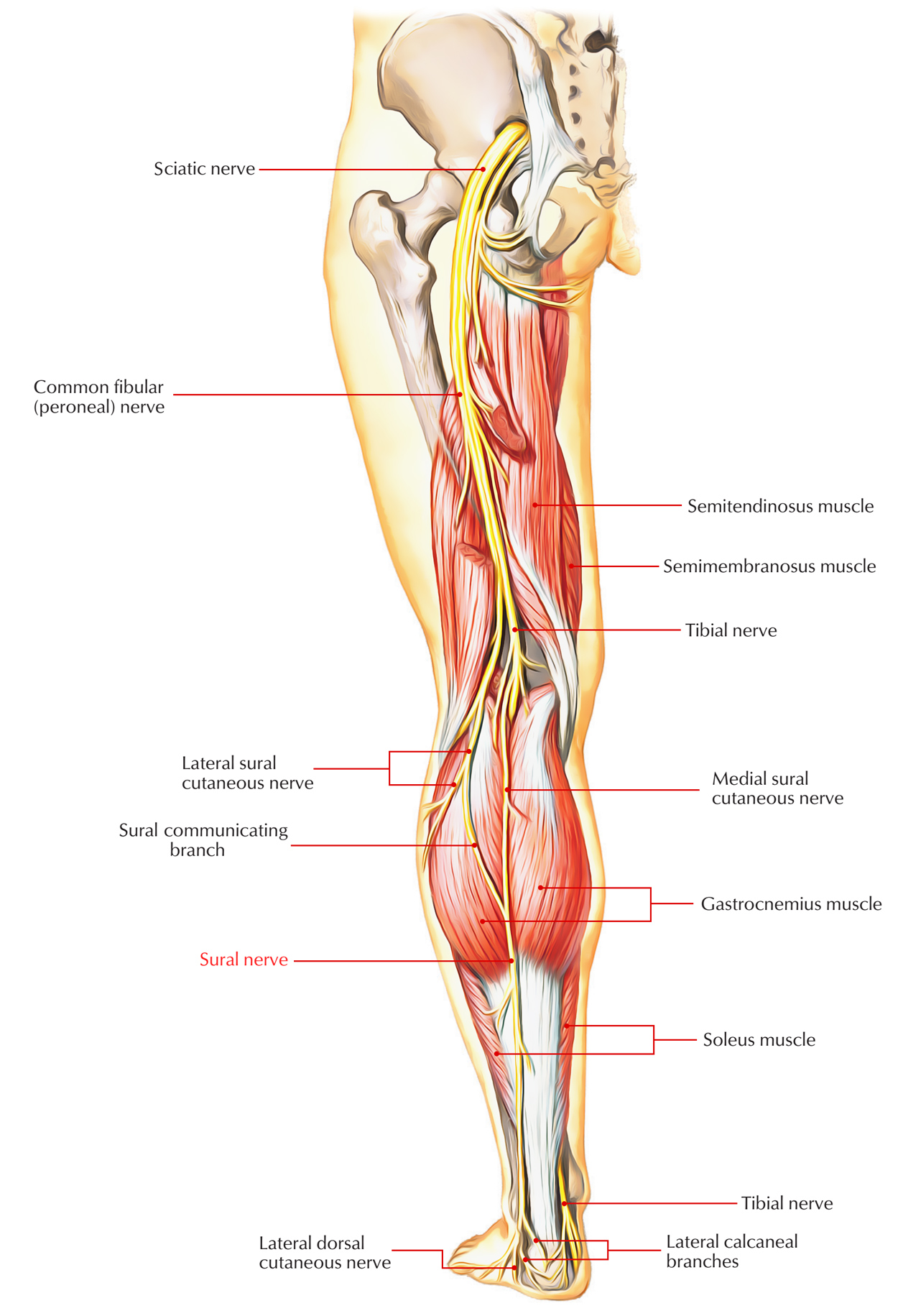
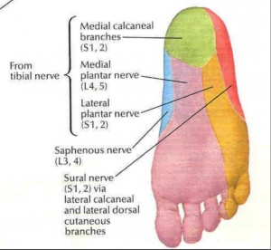
The sural nerve branches from the tibial and common fibular nerves and is responsible for feeling on the outside of the foot and the small toe.
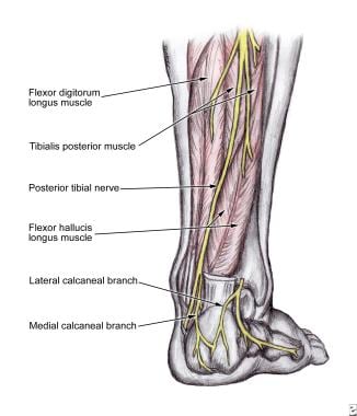
Nerve anatomy of foot. Medial calcaneal nerve innervates plantar medial heel. All of these nerves extend as branches of nerves in the leg that pass through the ankle and into the foot. Clinical anatomy for dummies.
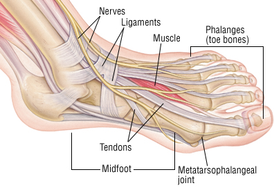
Terminal branches supply skin on the medial side of the proximal foot and enter the foot in superficial fascia on the medial part of the ankle. Pain swelling redness and bruising may be signs of a fracture. The anatomy of the nerves of the foot and ankle is complex and familiarity with the normal anatomy and course of these nerves as well as common anatomic variants is essential for correct identifi.
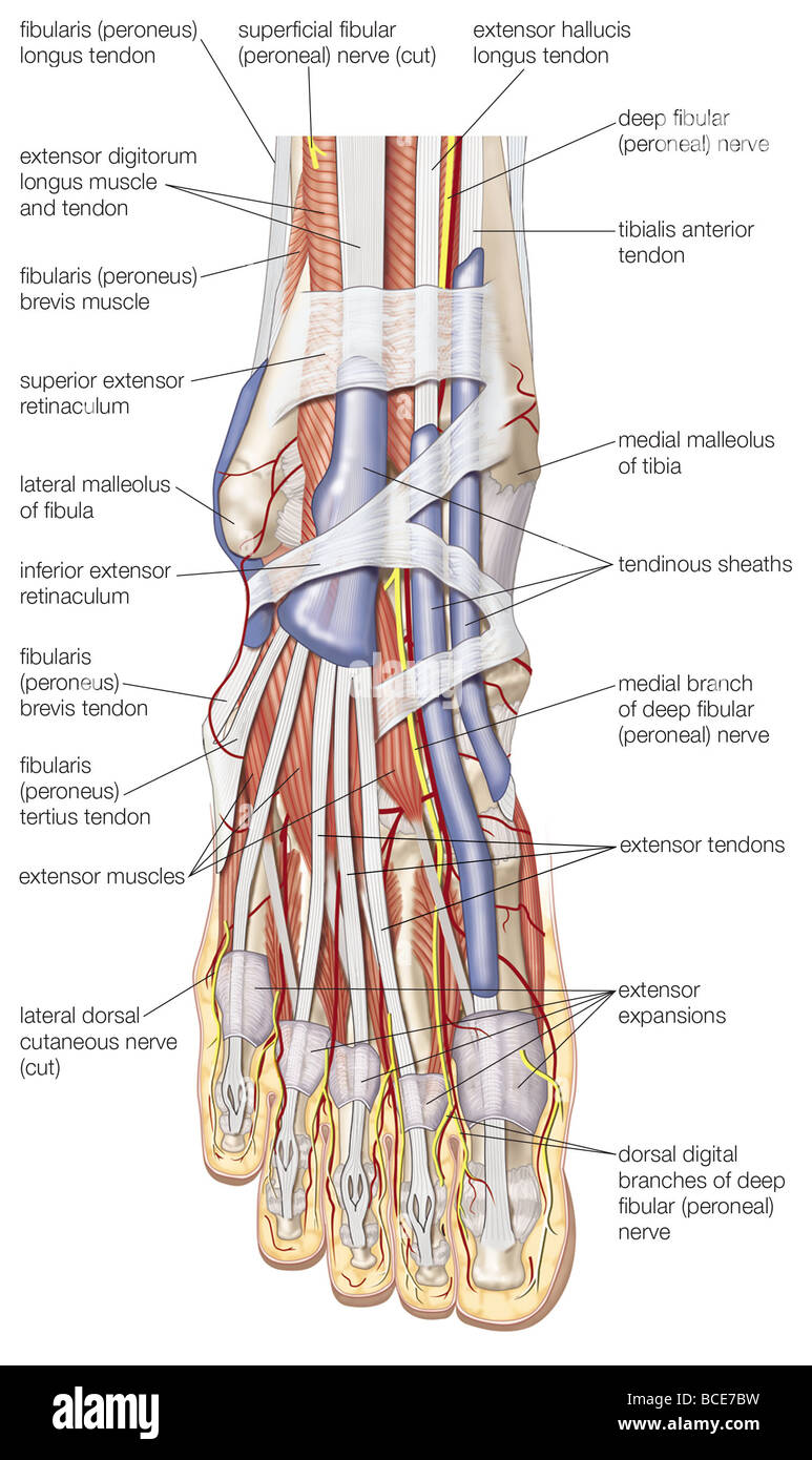
At risk proper branch of medial plantar nerve at risk with medial plantar approach to the tibial sesamoid. The nerves of the foot help move the body and keep balance both while its moving and at rest. The dorsal digital nerves are those nerves of the foot that cross the top surface and insert in the digits or toes.
Lumbircals to 2nd and 3rd toes. Many times an injured nerve will cause intense pain and heat felt within the foot. The sural nerve branches from the tibial and common fibular nerves and is responsible for feeling on the outside of the foot and the small toe.
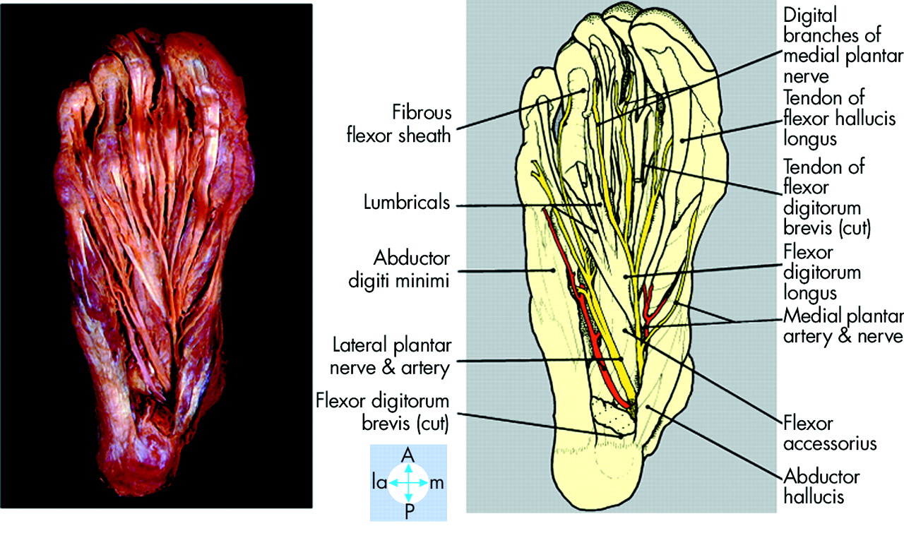
Medial plantar nerve innervates abductor hallucis. Saphenous nerve the saphenous nerve is a cutaneous branch of the femoral nerve that originates in the thigh. This nerve is a branch of the femoral nerve and runs down the medial portion of the leg to the medial part of the foot and innervates the skin on the medial side of the ankle and foot.
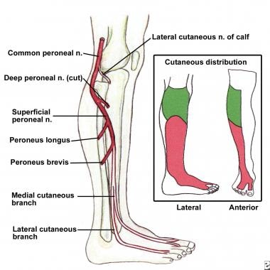
Nerves act as a network communicating important information from the foot to the brain. It innervates the skin on the lateral side of the leg and foot. A viral infection in the sole of the foot that can form a callus with a central dark spot.
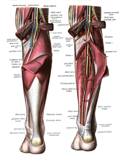
All of these nerves extend as branches of nerves in the leg that pass through the ankle and into the foot. The nerves of the foot help move the body and keep balance both while its moving and at rest. Branches of the tibial nerve.
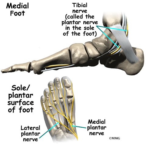
As such when the sciatic nerve is impaired it can lead to muscle weakness andor numbness or tingling in the leg ankle foot andor toes. On the sole the nerves of the foot are the medial and lateral plantar nerves which arise from the tibial nerve in the heel. The metatarsal bones are the most frequently broken bones in the feet either from injury or repetitive use.
 Lower Extremity Innervation Msk Medbullets Step 1
 Plantar Foot Anatomy Nerves Fa07 Foot Anatomy Human
 Nerve Blocks Of The Foot And Ankle Down East Emergency
 Common Peroneal Nerve Neurologyneeds Com
 Dorsal View Of The Right Foot Showing The Major Muscles
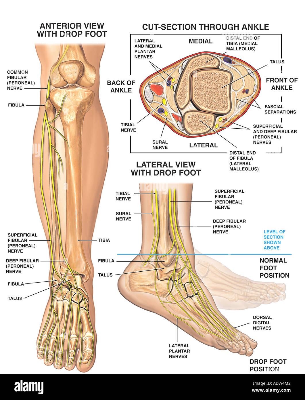 Anatomy Of The Foot And Ankle With Foot Drop Deformity Stock
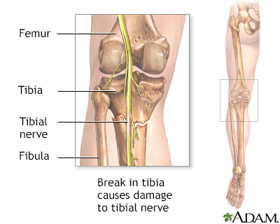
 What Is The Anatomy Of The Tibial Nerve Relevant To A
 Tibial Nerve Physiopedia
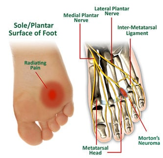
Morton S Neuroma Physiopedia
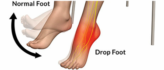
 Foot Drop Background Anatomy Pathophysiology
 Ankle Block Landmarks And Nerve Stimulator Technique Nysora
 Anatomy Foot Lower Leg Physician Assistant 2014 With
 Nerves Of Foot Earth S Lab
 The Foot And Ankle Practical Office Orthopedics
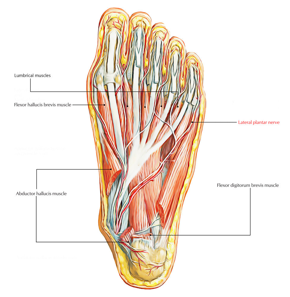 Easy Notes On Lateral Plantar Nerve Learn In Just 3
 Figure 1 From Ultrasound Guided Ankle Blocks A Review Of
 Chronic Heel Pain A Case Of Baxter S Nerve Injury
 Tibial Nerve Dysfunction Information Mount Sinai New York
![]() Lower Extremities Arteries And Nerves Anatomy Branches
Lower Extremities Arteries And Nerves Anatomy Branches
 Ankle Block Landmarks And Nerve Stimulator Technique Nysora
 Lateral Plantar Nerve Injury Following Steroid Injection For
 Core Anatomy Winding Round To Foot Drop Which Nerve Is
 Foot Anatomy Eorthopod Com
 Ulnar Nerve Anatomy Orthobullets
 Chapter 38 Foot The Big Picture Gross Anatomy
 Pin On Health
 Ankle Foot Anatomy
 Foot Nerve Anatomy Stock Photos Page 1 Masterfile
 Nerve Foot Images Stock Photos Vectors Shutterstock
 Foot Sprain Harvard Health
 Medial Plantar Nerve Physiopedia
Second Toe Flap
 Tarsal Tunnel Syndrome Foot Ankle Orthobullets


Posting Komentar
Posting Komentar