For a rat brain larger forceps are. Access online via elsevier 2006.
 Limbic System Wikipedia
The laboratory rat was developed from the norwegian rat rattus norvegicus by an american physiologist henry donaldson who started a breeding colony in 1906 at the wistar institute in philadelphia.

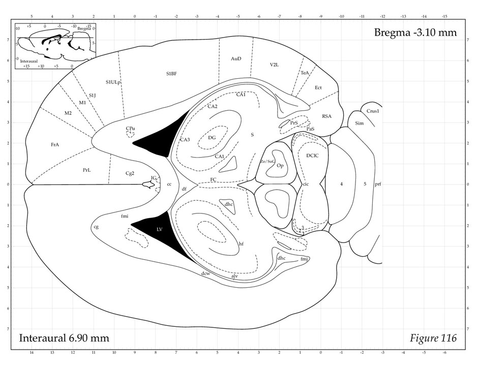
Rat brain anatomy. The rat brain is analyzed through stereotaxic localization of discrete brain areas and the subdivisions of many areas of rat brain are mapped using plates and diagrams. Developed at the wistar institute. Paxinos george and charles watsonthe rat brain in stereotaxic coordinates.
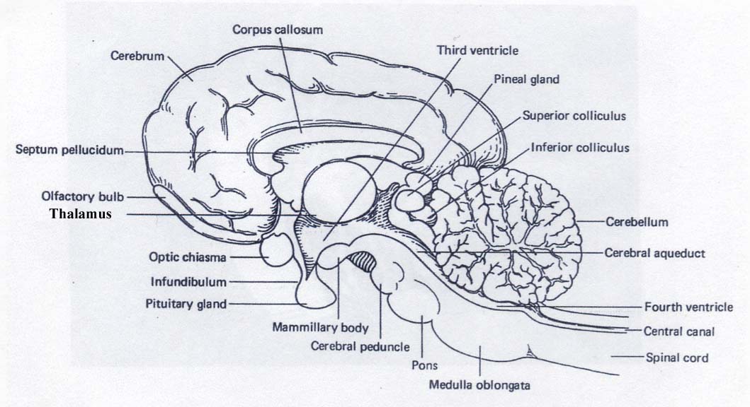
In the r emaining brain the genus corpus callosum gcc. The medical scheme focuses on the layout of the adult brain and names regions based on location and functionality. 139 and 142b is now well visible as.
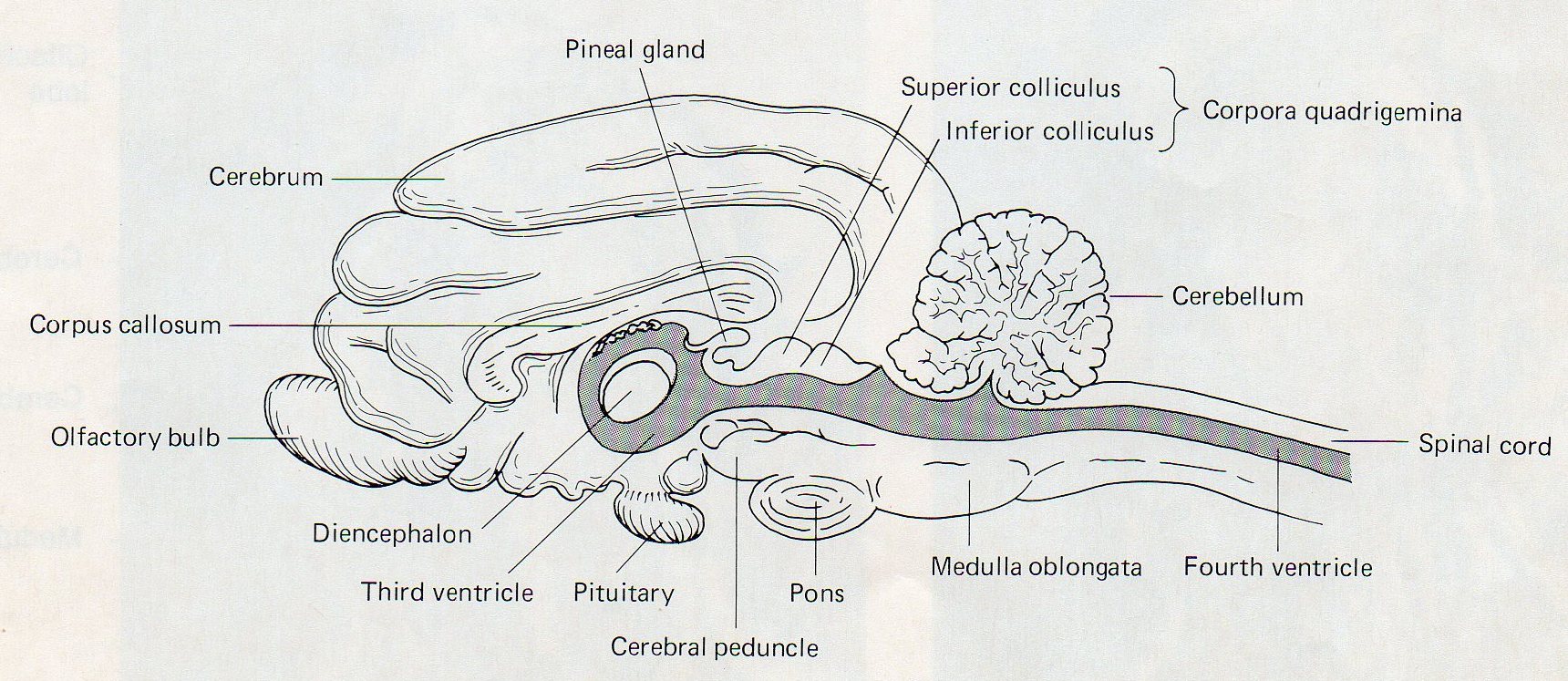
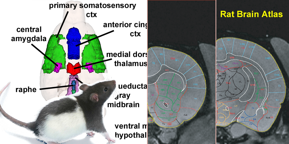
Rat atlas the rat atlas is a three dimensional 3d computerized map of rat brain anatomy created with digital imaging techniques. The anatomy of the brain is often discussed in terms of either the embryonic scheme or the medical scheme. Rat brain pictures dorsal aspect of brain and rostral two ventral aspect of the brain and junction of segments of spinal cord.
Dissection of rodent brain regions. Three principal strains are now commonly used for scientific study. The is based on high resolution isotropic ex vivo t2 weighted mri and dti data acquired at the duke center for in vivo microscopy at resolutions of 39 μm and 78 μm respectively.
Open arr ow figs. The 3 d rat brain atlas currently includes 60 structures while 30 structures have been incorporated in the 3 d mouse brain atlas. The structures included are the outer boundaries of the brain and areas zones and nuclei of the cerebral cortex hippocampus basal ganglia thalamus amygdala as well as major fiber tracts figures figures2a 2 a a2c 2 c c2e 2 e and and2g.
It was first presented as a poster and demo session at the incf booth of sfn 2009 in chicago. The specimen is an 80 day old male sprague dawley rat. A tool by matt gaidicamatt gaidica.
Development the scalable brain atlas is developed by rembrandt bakker in collaboration with many othersit uses exploratory work of gleb bezgin creator of the cocomac paxinos3d. The waxholm rat atlas is an open access volumetric atlas of the sprague dawley rat brain. Electronic sharing and interactive use are benefits afforded by a digital format but the foremost advantage of this 3d map is its whole brain integrated representation of rat in situ neuroanatomy.
The embryonic scheme focuses on developmental pathways and names regions based on embryonic origins. Photographs of sufficient magnification are included to permit investigators to judge for themselves the veracity of the atlas delineations. Medulla with spinal cord.
 Figure 4 9 The Distribution Of Opioid Receptors In The Rat Brain
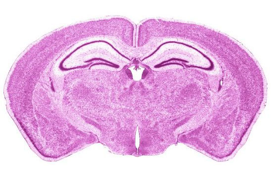
 Rat Brain Coronal At Ac A Photo On Flickriver
 Neuroanatomy Wikipedia
 Rat Brain Anatomy
 Rat Brain Anatomy Slk Art
 Image Of Seven Transverse Coronal Sections In The Brain
 1 Anatomy Of The Hippocampal Formation A Schematic Rat
 Nervous System Sciencedirect
Secrets Of The Brain
 Physiological Psychology
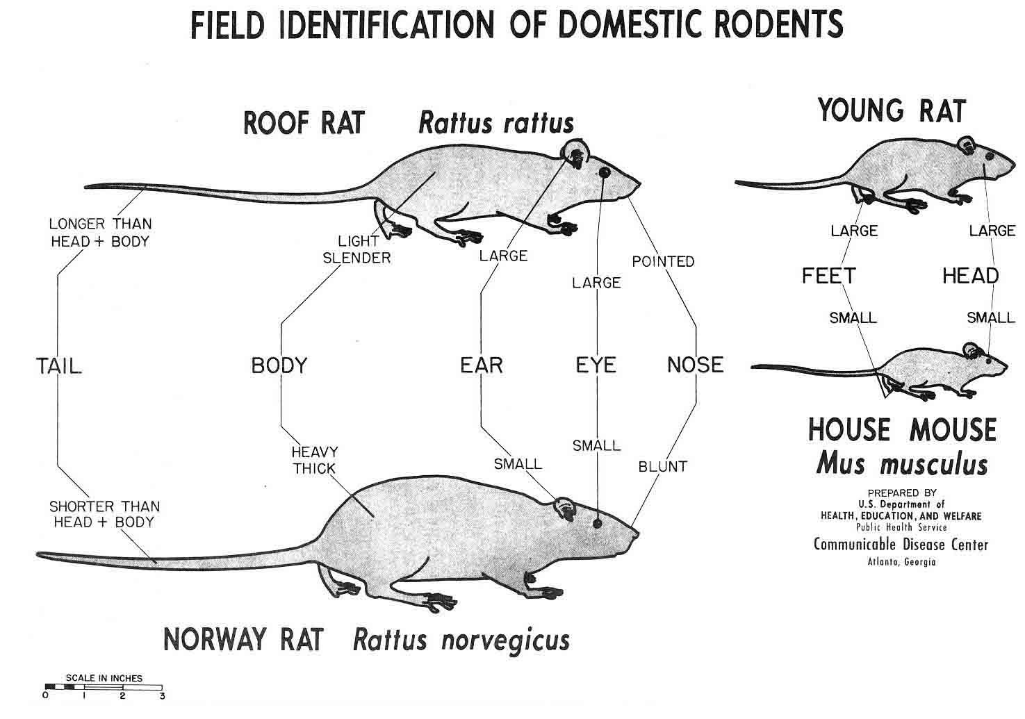
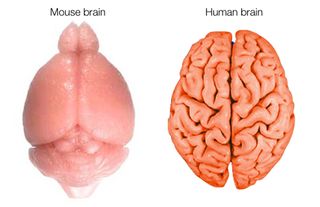
 How Can Studies On Rats Apply To Humans Howstuffworks
 Gross Neuro Anatomy
 Voltage Gated K Channel B Subunits Expression And
 Rat Brain Anatomy Artwork Stock Image C020 6714
 Physiological Psychology
Rat Brain Pictures
 Rat Brain Comparative Anatomy

 Figure 1 From Inhibitory Interneurons In The Piriform Cortex
 Rat Brain Anatomy
 Stereotaxic Surgery For Excitotoxic Lesion Of Specific Brain

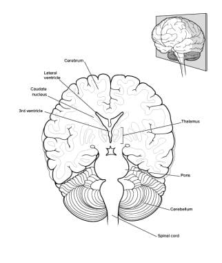 Brain Anatomy Overview Gross Anatomy Cerebrum Gross
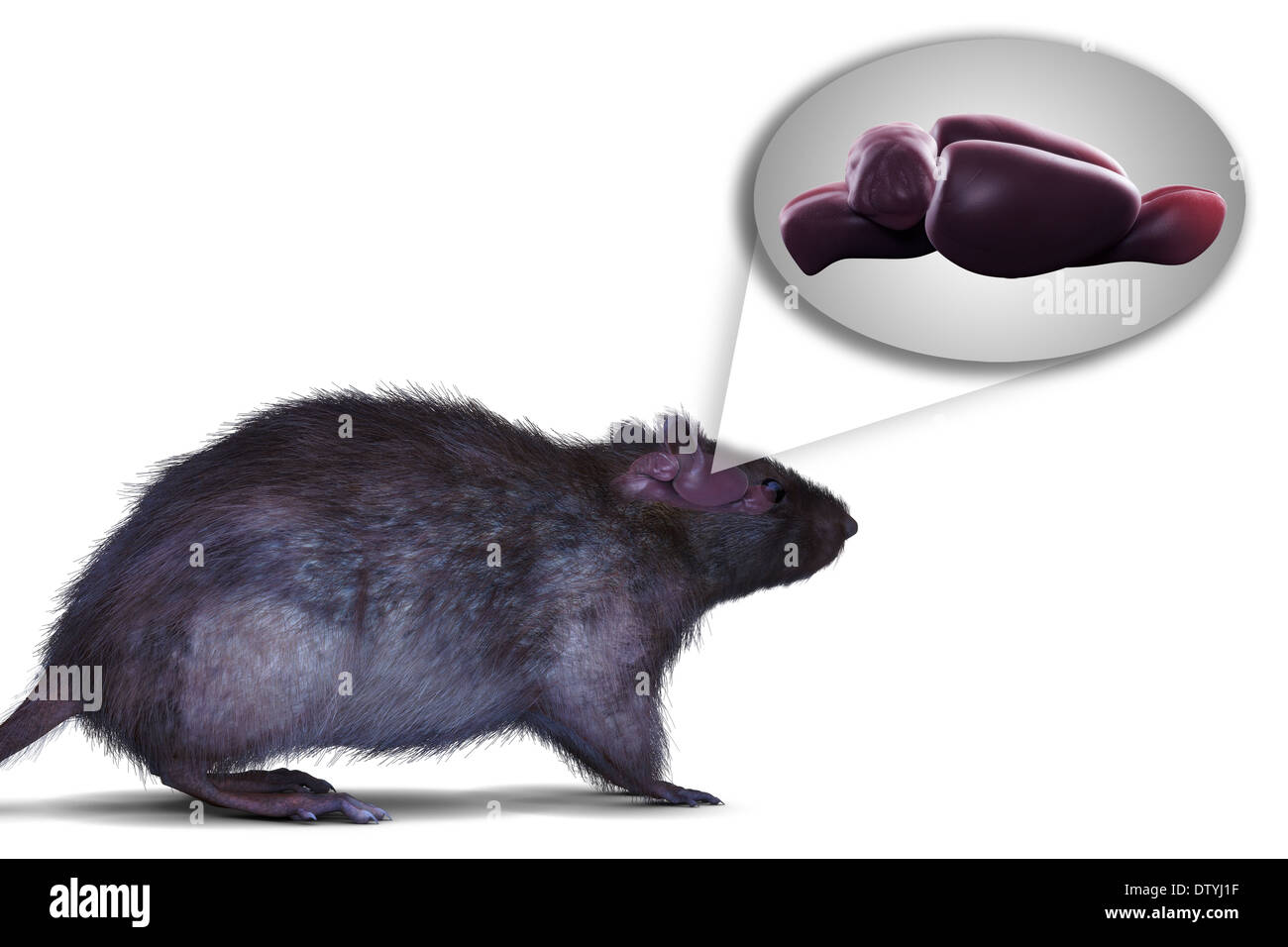
 All About Rats

 Schematic Representation Of Gross Anatomical Brain Areas Of
 Rat Brain Anatomy Stock Photo 66989707 Alamy
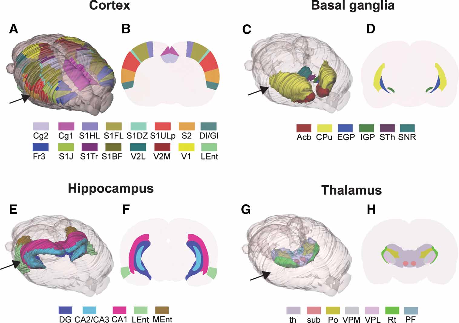 Frontiers Three Dimensional Atlas System For Mouse And Rat
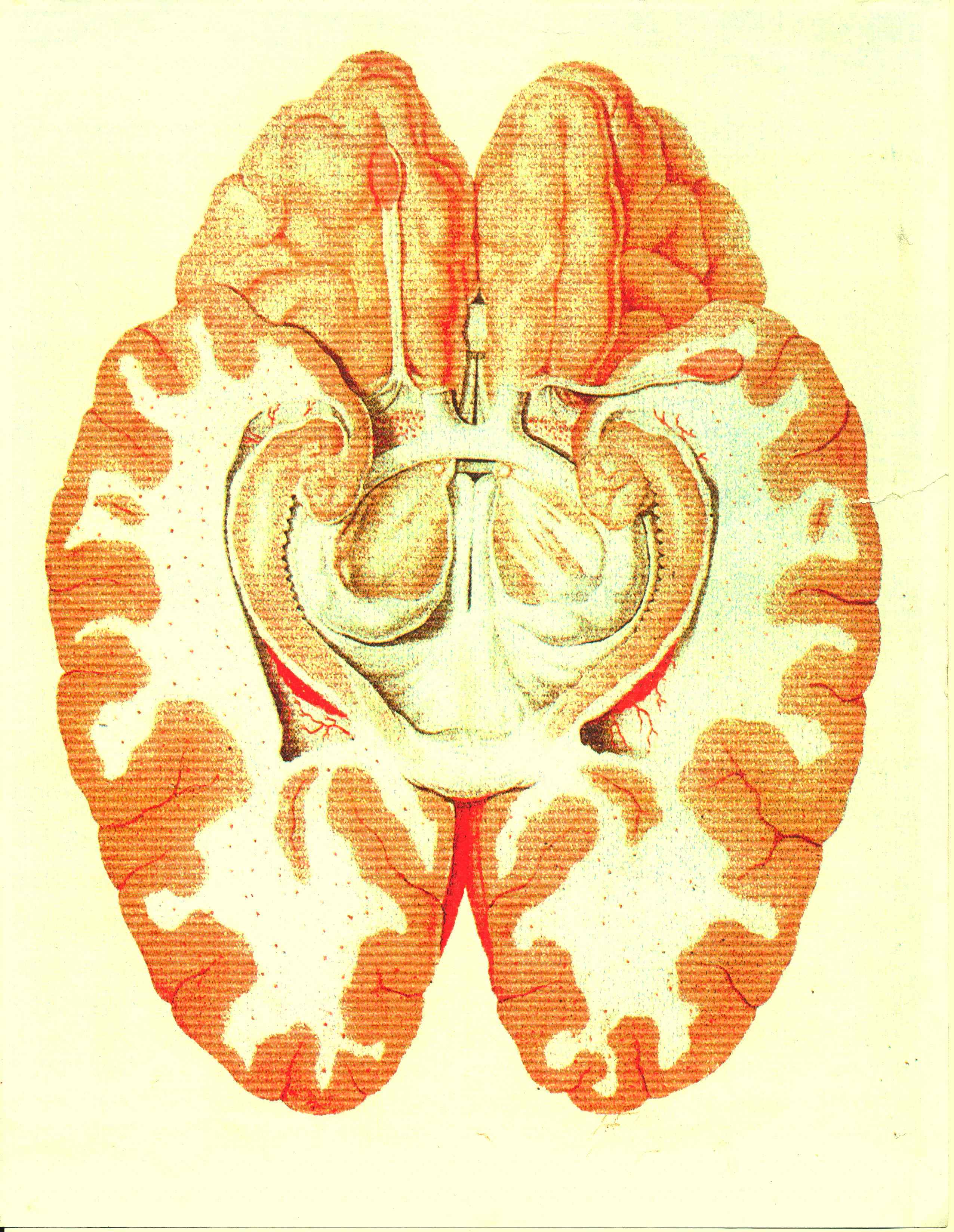
 A Vascular Brain Anatomy Of The Rat Reproduced From 10
 How The Human Brain Gets Its Wrinkles Live Science


Posting Komentar
Posting Komentar