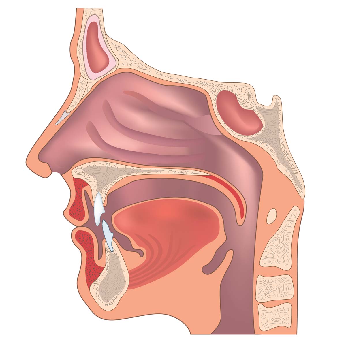
Histological changes of a sample can be then checked under the microscope. The shaded area represents the region of the nasopharynx.
Basic mouth and throat anatomy for anesthesia and ent rotations.

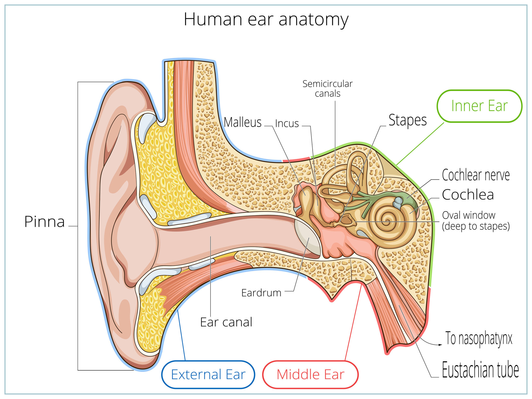
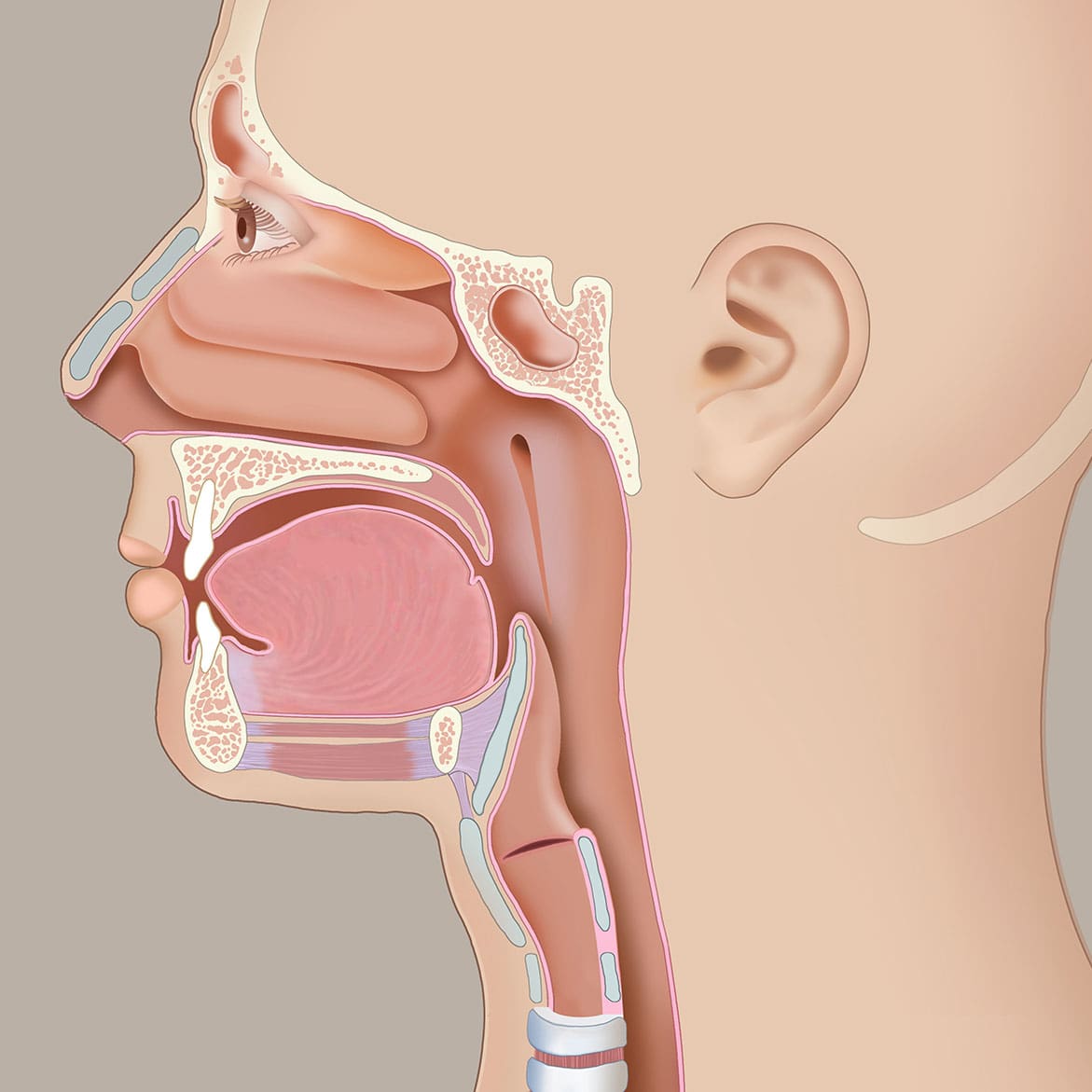
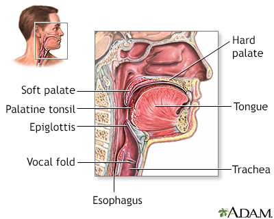
Anatomy of ear and throat. Throat begins at the back of your mouth and ends in the upper part of the stomach. The throat is the muscular and cartilage tube that acts as the passageway for air food and liquid and also helps in forming speech. Accordingly throat is a narrow elongated part connecting your head to the shoulders.
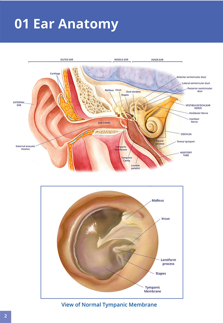
Human anatomy models of the nose and sinuses are perfect for working with students or patients with allergies and many of our larynx models feature functional vocal cords. The throat consists of. The nerves that take sound to the brain are found in the inner ear.
Adenoids is the term given to the lymphatic tissue collection. Roof and posterior wall. Individual ear anatomy models range from desk sized models to giant sized ears featuring removable bones and internal organs.
They are made up of lymph tissue and are located at the back and the sides. Each of these parts in the throat anatomy has been discussed in detail in the following sections. The throat consists of the larynx responsible for producing sound these muscles also allow food to pass down into the oesophagus.
A flap of soft tissue located just above the vocal cords. Different parts of the throat. The dark like at the front edge of the neck which shows where the throat is.
Throat parts pictures functions. The larynx is a cylindrical grouping of cartilage muscles. There are two major sections of the throat they are the pharynx and the larynx.
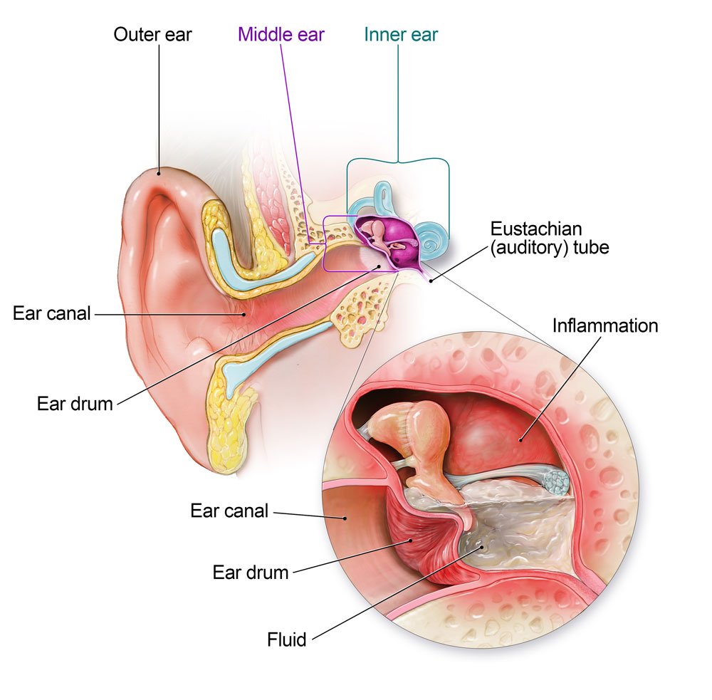
Ear infections in adults and kids adults and children get ear infections for the same reasons. They imperceptibly merge with each other. The human throat anatomy can mainly consists of the following parts.
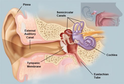
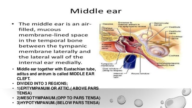
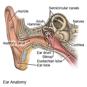
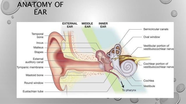
These structures are shaped in a ring or a muscular tube. Gross anatomy of throat. The middle ear includes the eustachian tube 3 which connects to the throat and the ossicles tiny bones 4 through which sound travels.
Anatomy of the nasopharynx. Your doctor for ear nose and throat ent can use laryngoscope to make a detailed investigation of the larynx and take samples biopsy of laryngeal mucosa. Larynx also known as the voice box.
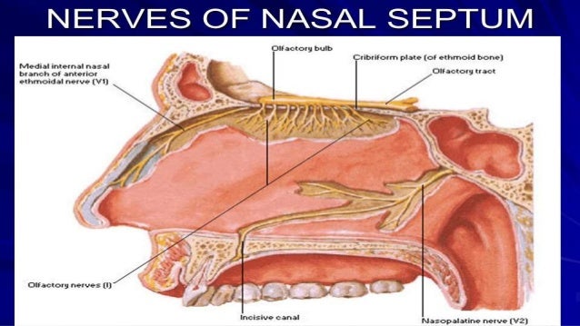
Ear nose and throat. As long as a person is alive throat functions non stop helping in breathing and swallowing. Anteriorly by the posterior end of the nasal septum and the posterior nasal apertures choanae.
 Common Ear Nose And Throat Complaints Everyday Health
 Picture Of The Ear Ear Conditions And Treatments
 It S All Connected Lateral Ent Health
 The Association Between Tinnitus The Neck And Tmj
 Figure Ear Anatomy Image Courtesy S Bhimji Md
 Anatomy And Physiology Of Ear Nose Throat And Newer
 Anatomy And Physiology Of Ear Nose Throat And Newer
 Ear Injuries Diving Everything You Need To Know Manta Dive
 Anatomy Of The Ear Professional Hearing Services
Anatomy And Physiology Of The Ear Children S Wisconsin
 Eustachian Tube Dysfunction What You Need To Know
 Ear Infection Community Antibiotic Use Cdc
 Struggling With Sinusitis Live Chat With An Ear Nose
 Demystifying The Ear Canal Consider Professional Ear
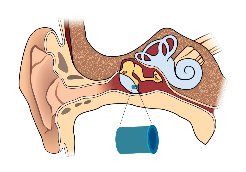 Ear Tubes Mississippi Ear Nose Throat Surgical Associates

 Emma Scheltema Illustration Visual Guide To Ear Nose
 29 Best Ent Ears Nose Throat Images Ear Anatomy Throat
 Ear Nose Throat Medical Quick Reference Guide
 Tragus Anatomy Britannica
 Tympanic Membrane
 Middle Ear An Overview Sciencedirect Topics
 Anatomy And Physiology Of Ear Nose Throat And Newer


Posting Komentar
Posting Komentar