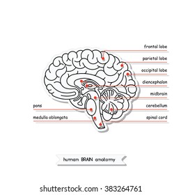
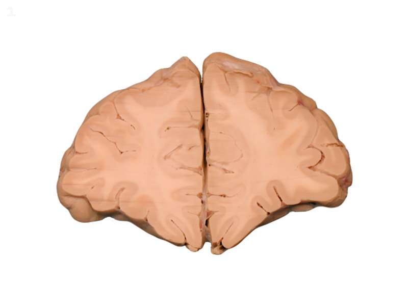
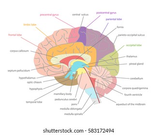
Frontal lobe medulla oblongata thalamus cerebellum hypothalamus and more. Cross section of brain cortex in this image you will find corpus callosum dorsomedial thalamic nucleus internal medullary lamina lateral thalamic nuclei the hypothalamus in it.
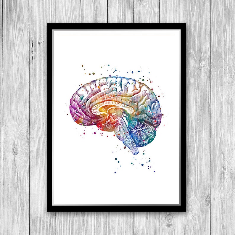 Neuroanatomy Art Brain Anatomy Cross Section Watercolor Print Medical Poster Doctor Office Decor Neurology
The cross sectional anatomy of the normal adrenal gland is identical on ct and mri.
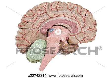
Cross section of brain anatomy. Helping your student understand the complexities of the human brain is easy with this realistically detailed cross section model. The right adrenal gland lies more cranially in the abdomen than the left adrenal. An mri was performed on a healthy subject with several acquisitions with different weightings.
Mri of the brain. You will also find stria medullaris caudate nucleus claustrum putamen globus pallidus amygdala optic tract as well. Spin echo t1 t2 and flair t2 gradient echo diffusion and t1 after gadolinium injection.
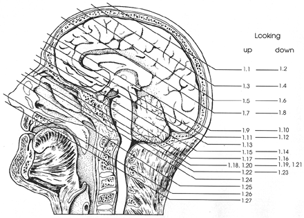
Is located in the floor of the third ventricle and is the master control of the autonomic system. Use the mouse scroll wheel to move the images up and down alternatively use the tiny arrows on both side of the image to move the images. Learn cross sections neuroanatomy brain with free interactive flashcards.
Cerebellum cross section of brain you are looking at a sagittalat a sagittal cross section of the brain cerebellum or little brain this structure controls movement posture and balance 2009 the university of texas health science center at san antonio this structure controls movement posture and balance. It plays a role in controlling behaviors such as hunger thirst sleep and sexual response. Made of durable soft foam this 2 piece brain features the main parts of the brain including.
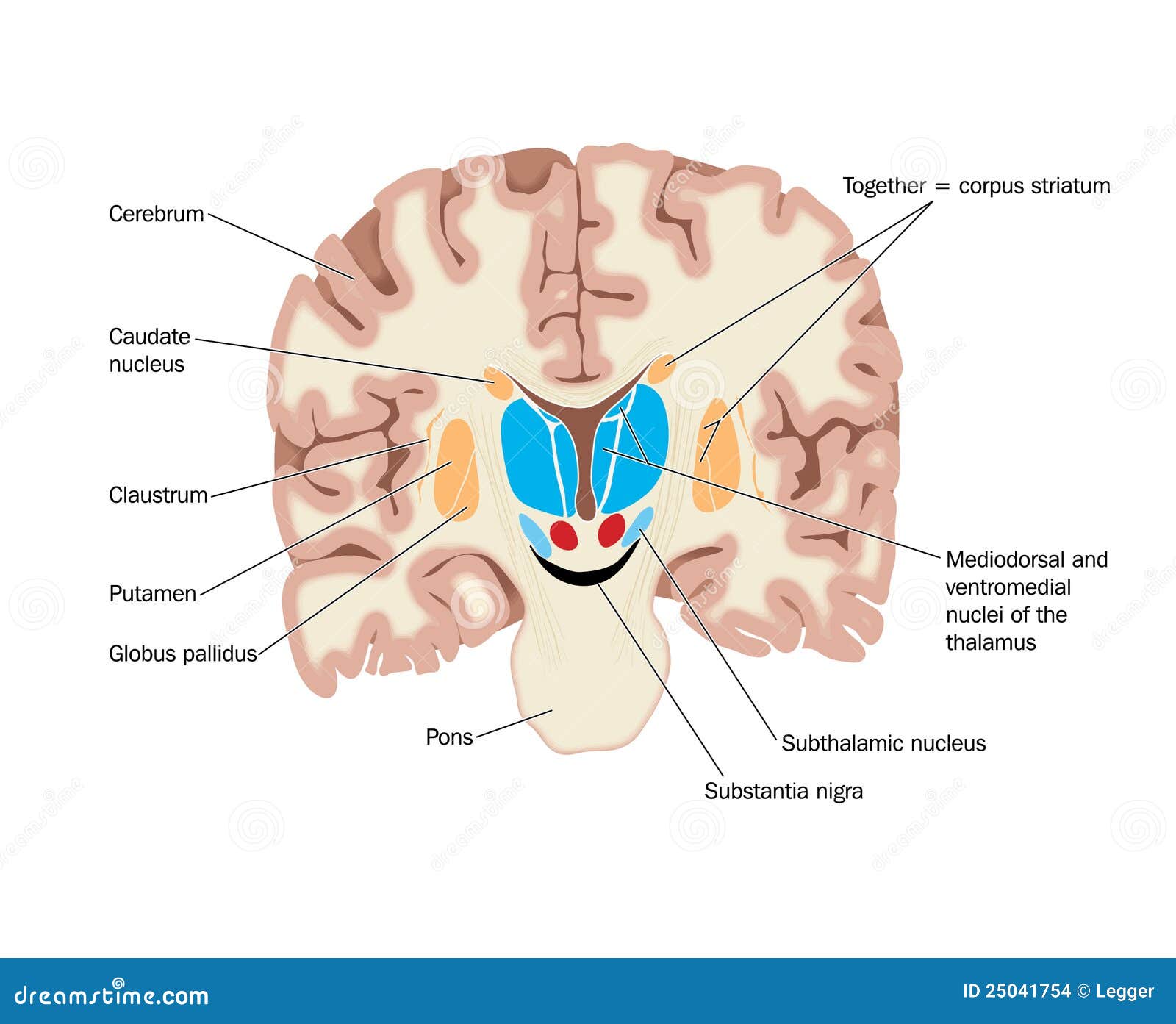
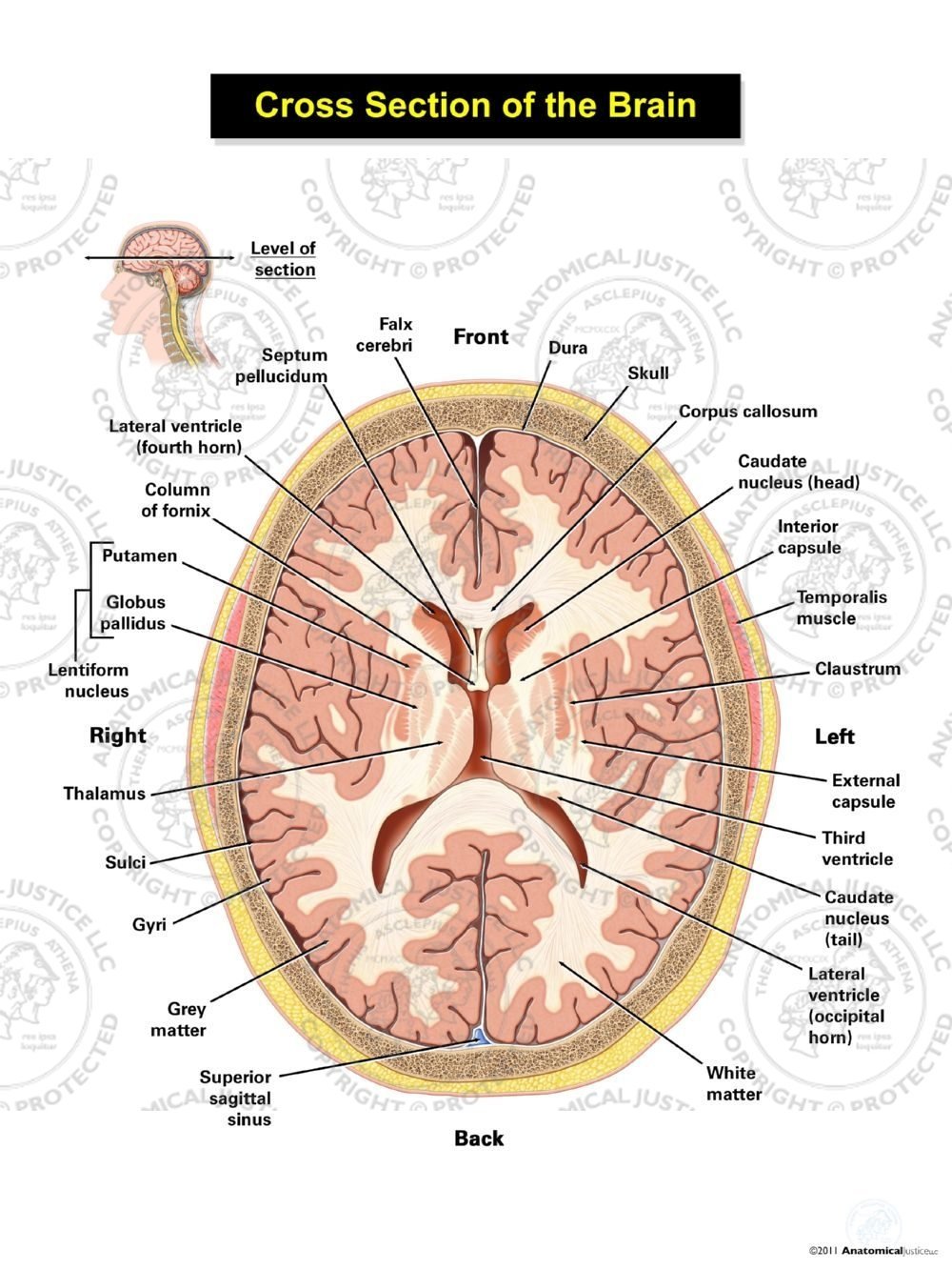
Choose from 500 different sets of cross sections neuroanatomy brain flashcards on quizlet. Coronal cross section showing the basal ganglia. It is superior to the upper pole of the right kidney whereas the left adrenal gland is anteromedial to the upper pole of the left kidney.
This mri brain cross sectional anatomy tool is absolutely free to use. We obtained 24 axial slices of the normal brain. Data and dicom images archived on our pacs.
 Brain Anatomy Cross Section Two Tone Mug Spreadshirt
 Brain Anatomy Cross Section 2
![]() Horizontal Sections Of The Brain Anatomy Kenhub
Horizontal Sections Of The Brain Anatomy Kenhub
 Human Skull Cross Section With Brain Stock Illustration
 Brain Anatomy Cross Section X By Erzebet S
 Anatomy Atlases Atlas Of Human Anatomy In Cross Section
 Cross Section Of The Brain Showing Nuclei Stock Vector
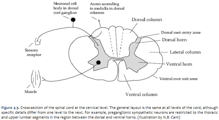 Duke Neurosciences Lab 2 Spinal Cord Brainstem Surface

 Cross Section Of Brain Images Stock Photos Vectors
 Man Head With Skull Cross Section With Brain Stock
 Colorful Cross Section Of Human Brain Anatomy Stock Photo
 Color Illustration Coronal Cross Section Showing The Basal

 Human Skull Mid Sagittal Cross Section With Brain Perspective

 Anatomical Model Of Human Brain Cross Section Picture
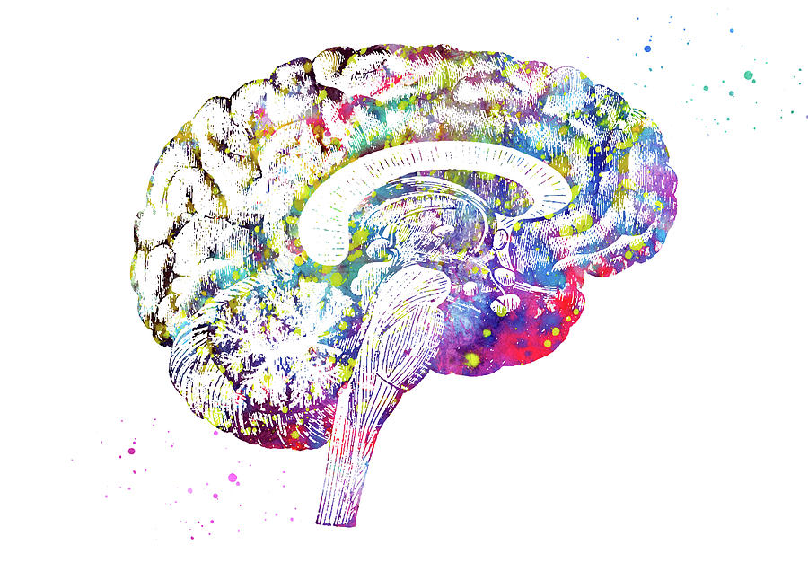
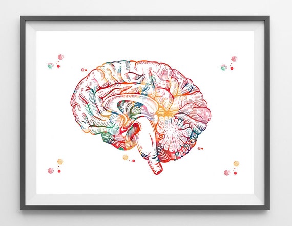 Brain Anatomy Cross Section Watercolor Print Human Brain Sagittal View Limbic System Poster Medical Art Neurology Illustration Anatomy Art
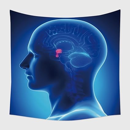
Amazon Com Home Decor Tapestry Wall Hanging Brain Anatomy
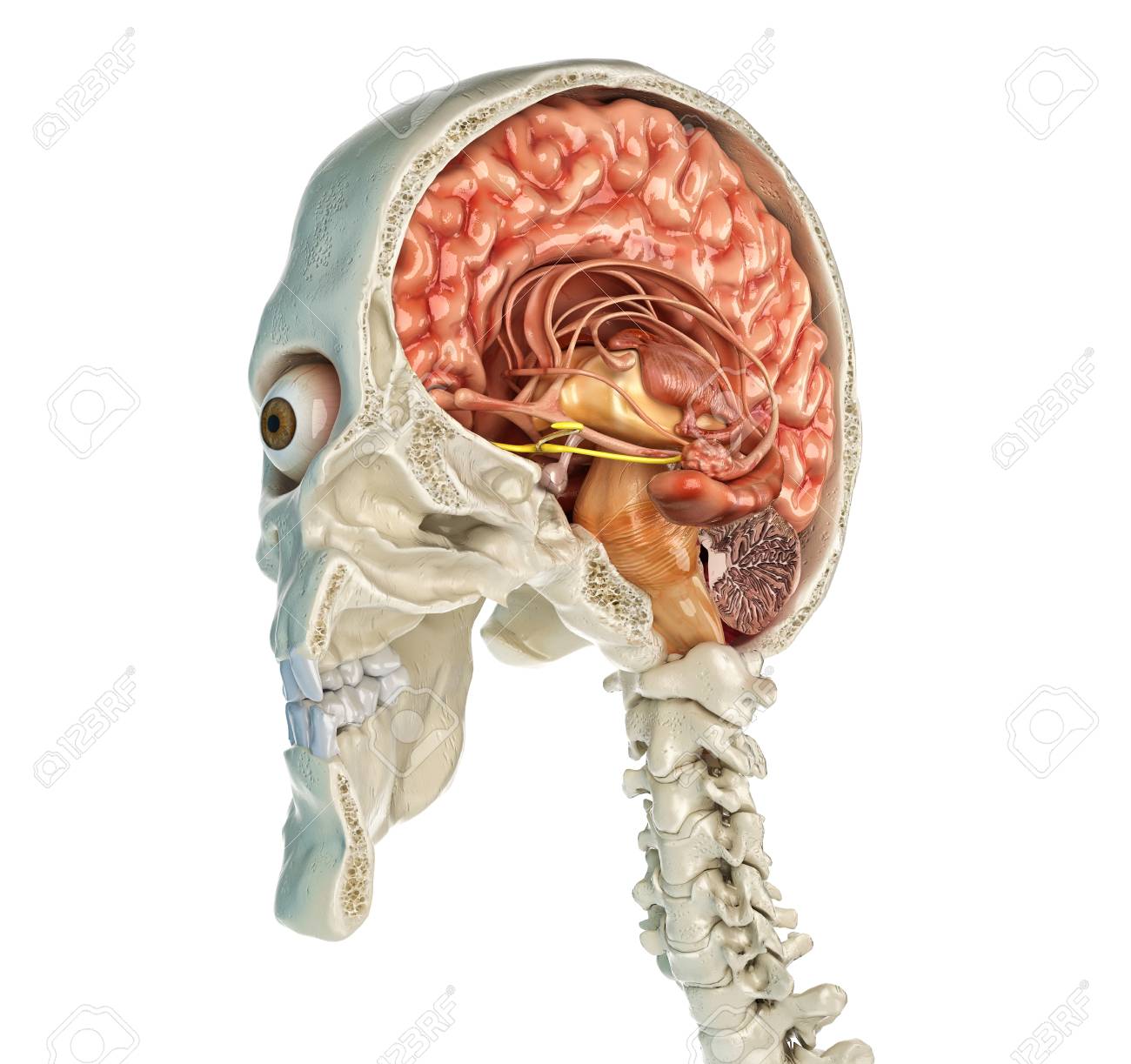
 Anatomy Head Cross Sections Model Brain Eye Ear Nasal
 The Cerebrum And Cerebral Hemispheres

 Arrangement Of Thalamic Nuclei A Cross Section Of The
 Cross Section Of The Brain Anatomy Dry Erase Sticky Wall Chart
 Cross Section Brain Images Stock Photos Vectors
 Human Brain Cross Section Anatomy Stock Illustrations 342
 Cross Section Of The Brain Ventricular Level


Posting Komentar
Posting Komentar