Anatomy of the abdomen and male pelvis using cross sectional imaging ct interactive atlas of human anatomy we have created an anatomical atlas of abdominal and pelvic ct which is an interactive tool for studying the conventional anatomy of the normal structures based on a multidetector computed tomography. The pelvic floor is primarily made up of thick skeletal muscles along with nearby ligaments and their investing fascia.
 Ct Anatomy Of The Pelvis
It is a basin shaped muscular diaphragm that helps to support the visceral contents of the pelvis.

Pelvic muscles anatomy ct. The main focus of this article will be the pelvic floor muscleson that topic there are several important questions that need to be answered. This is the first part of a two part tutorial on the pelvic floor and discusses the muscles which make up the pelvic diaphragm. This mri male pelvis axial cross sectional anatomy tool is absolutely free to use.
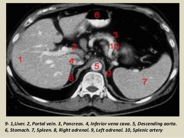
2 psoas muscle 3 lumbar vertebra 12 kidney 15 liver 17 stomach 19 gall bladder 22 small bowel. Talos i f jakab m kikinis r. They support the pelvic organs especially during increases in intra abdominal pressure and also aid in urinary and faecal continence.
The muscles of the pelvis form its floor. The following structures are discussed. Pelvic muscles ct anatomy and ct scan of the abdomen and pelvis shows a normal appendix 7 pelvic muscles ct anatomy pelvic muscles ct anatomy and ct scan of the abdomen and pelvis shows a normal appendix gallery at human diagram chart.
Learn the diagnosis of ct and methods of computed tomography. There are many muscles that form the pelvic floor including puborectalis pubococcygeus iliococcygeus and coccygeus. Atlas of ct anatomy of the abdomen.
15 liver 16 oesophagus 17 stomach. Anatomy ct axial abdomen and pelvis male male abdomen and pelvis ct scan form no 1. Ct anatomy of the pelvis.
This photo gallery presents the anatomy of the abdomen by means of ct axial coronal and sagittal reconstructions. Use the mouse scroll wheel to move the images up and down alternatively use the tiny arrows on both side of the image to move the images on both side of the image to move the images.
 Abdomen And Pelvis Ct
 Abdomen And Pelvis Anatomy Of The Dog On Ct
 Ecr 2014 C 0356 The Pelvis Revisited A Pictorial Review

 The Male Pelvis Mr Anatomy Atlas Of The Prostate Bladder
 Sectional Anatomy Of Abdomen
 The Ultimate Pelvic Anatomy Resource Pelvic Guru Featured
 Pelvic Ct Angiography Showing The Anatomy And Nomenclature
 Pelvis An Overview Sciencedirect Topics
 Above Shows A Number Of Possible Measurements Using Mri
 The Ct Anatomy Tutor
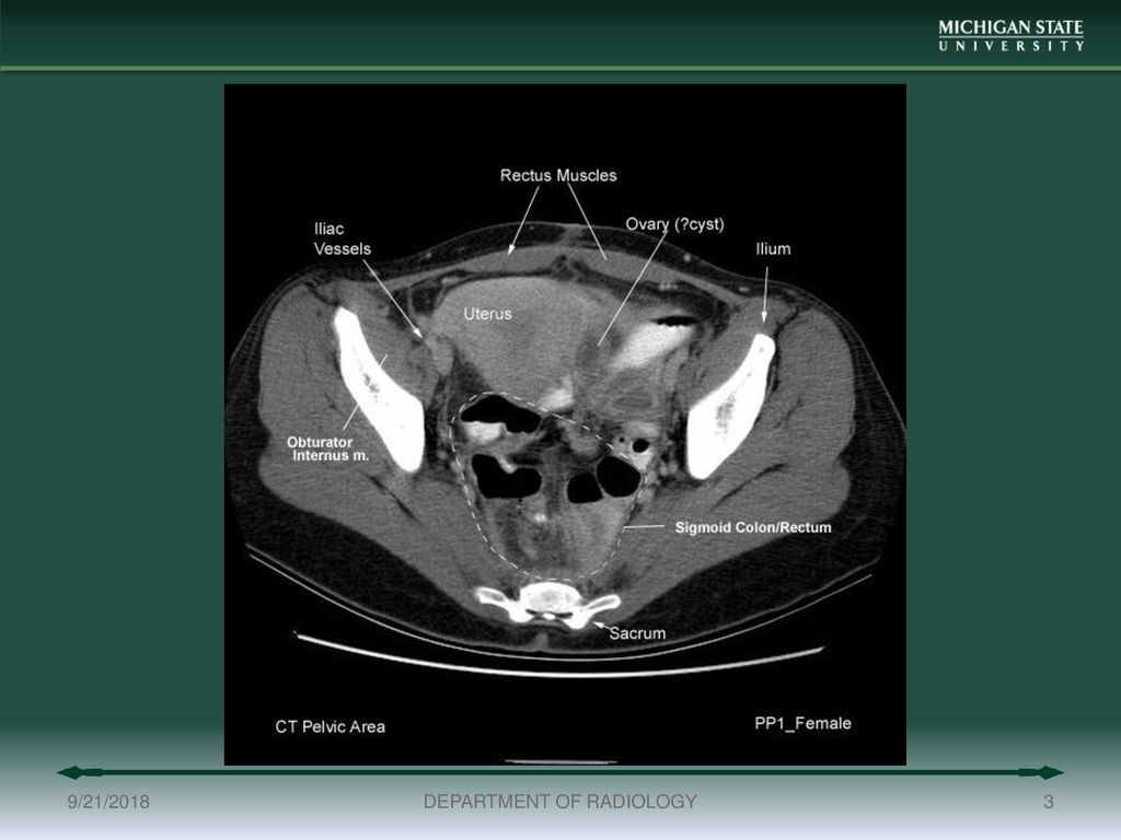
 Body Ct Modules Ct Of The Ovaries And Uterus Ppt Download
 Abdomen And Pelvis Ct

 Abdomen And Pelvis Ct
 Abdomen And Pelvis Ct
 Pelvis Perineum Anatomy Ppt Download
 The Pelvis Basicmedical Key
 Pelvis Wikipedia
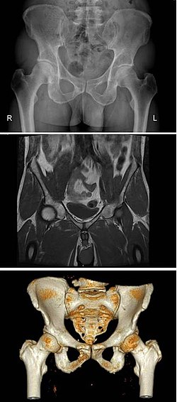
![]() Medical Imaging And Radiological Anatomy X Ray Ct Mri
Medical Imaging And Radiological Anatomy X Ray Ct Mri
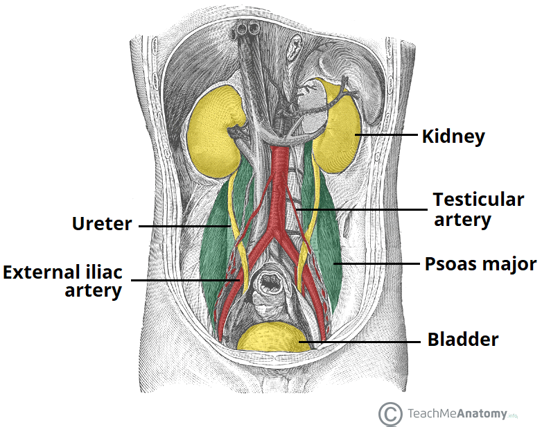 The Ureters Anatomical Course Neurovascular Supply
The Ultimate Pelvic Anatomy Resource Pelvic Guru Featured
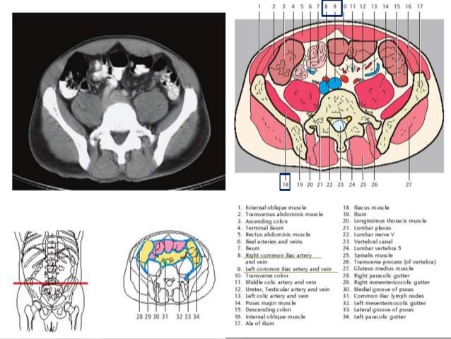
 Presentation1 Pptx Ct Normal Anatomy Of The Abdomen And Pelvis
 Abdominopelvic Cavity And Peritoneum On A Ct
 Startradiology
 The Pelvis Ct Anatomy Mp4


Posting Komentar
Posting Komentar