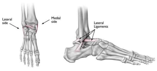
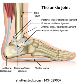
Foot ankle anatomy muscles tendons and ligaments. Posterior talofibular spans between the lateral malleolus and the posterior aspect of the talus.
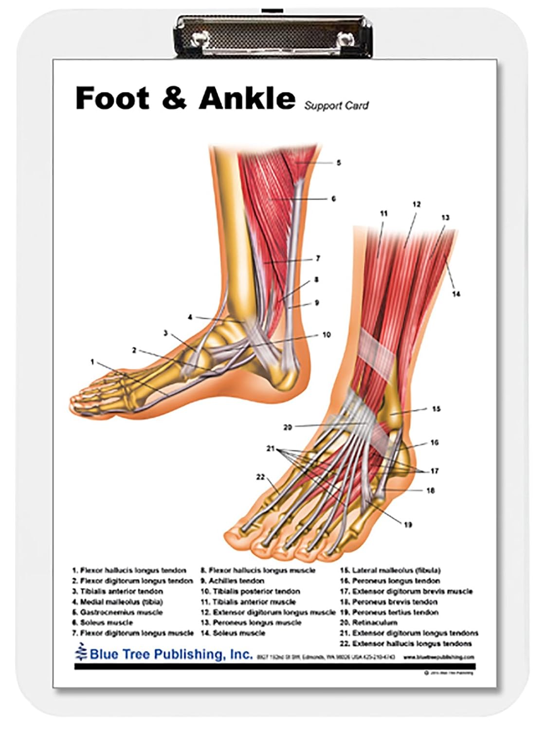 Amazon Com Foot Ankle Insert Clipboard Industrial
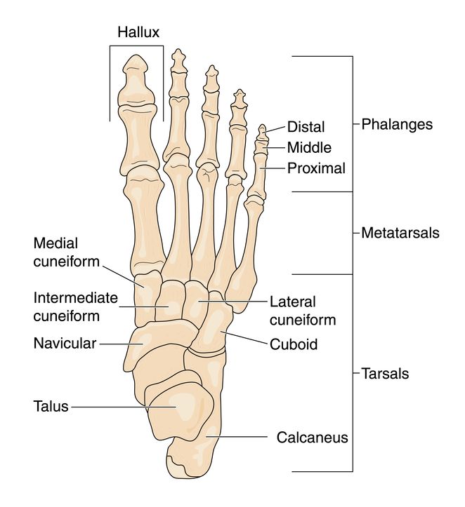
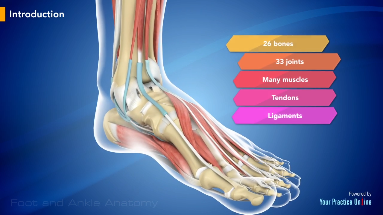
The foot consists of thirty three bones twenty six joints and over a hundred muscles ligaments and tendons.
Anatomy of the ankle. It resists over inversion of the foot and is comprised of three distinct and separate ligaments. Anterior talofibular spans between the lateral malleolus and lateral aspect of the talus. Biomechanically a certain amount of motion is allowed in all planes with respect to the distal ends of the tibia and fibula.
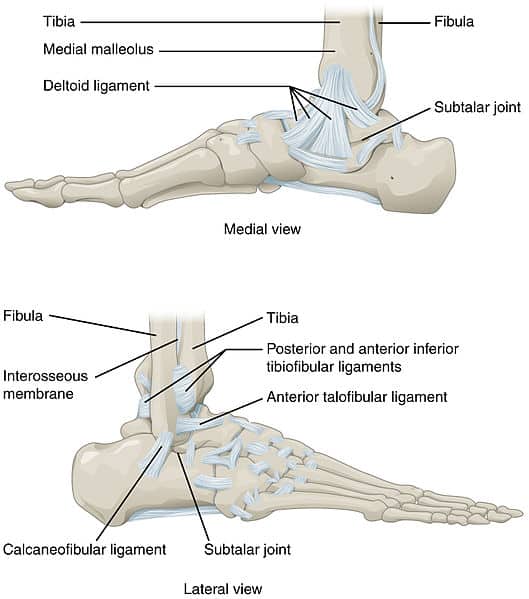
The ligaments around the ankle can be divided depending on their anatomic position into three groups. The shin bone tibia. Tendons are elastic tissues made up of collagen.
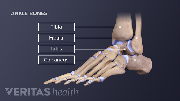
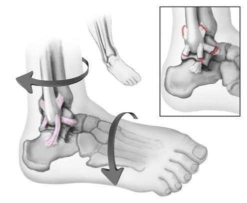
What we normally think of as the ankle is actually made up of two joints. The ankle is the joint between the foot and leg composed of three separate bones. Medically reviewed by healthline medical team on april 8 2015.
There are elastic tissues tendons in the foot that connect the muscles to the bones and joints. The outer bone is the fibula or calf bone. The largest and strongest tendon of the foot is the achilles tendon which extends from the calf muscle to the heel.
Soft tissues of the foot and ankle ligaments. The subtalar joint and the true ankle joint. Anatomy of the ankle the ankle is a complex mechanism.
The tibia and fibula are connected throughout their length by an interosseous membrane. Foot and ankle anatomy is quite complex. The syndesmosis of the ankle refers to the membrane connecting the tibia to the fibula.
A foot bone that sits above the heel bone talus. Its strength and joint function facilitate running jumping walking up stairs and raising the body onto the toes. These all work together to bear weight allow movement and provide a stable base for us to stand and move on.
Ligaments are strong dense and flexible bands of fibrous connective tissue. There are many muscles that help to move and support the ankle and foot. The inner bone is the tibia or shinbone which supports most of a persons weight when standing.
The ankle is a large joint made up of three bones. Fascia is a broad fibrous. The thinner bone running next to the shin bone fibula.
The lateral ligaments the deltoid ligament on the medial side and the ligaments of the tibiofibular syndesmosis that join the distal epiphyses of the bones of the leg tibia and fibula.
 Ankle Foot Atlas Of Anatomy
 Ankle Bones Anatomy Ankle Anatomy Foot Anatomy Ankle
 Anatomy Of An Ankle Sprain Bouldercentre For Orthopedics
![]() Ankle Joint Anatomy Bones Ligaments And Movements Kenhub
Ankle Joint Anatomy Bones Ligaments And Movements Kenhub

 Ankle Foot Atlas Of Anatomy

:max_bytes(150000):strip_icc()/GettyImages-687795661-969f111662dc4df7a9c3d78c17045ea6.jpg) Anatomy And Physiology Of The Ankle For Sports Medicine
Anatomy And Physiology Of The Ankle For Sports Medicine
 Ankle Wikipedia
 Foot Bones Anatomy Conditions And More
 Applied Surgical Anatomy Of The Approaches To The Ankle

 Ankle Joint Anatomy And Osteoarthritis
 Ankle Images Stock Photos Vectors Shutterstock

 Anatomy Of The Foot Ankle
 Ankle Foot Anatomy
 Doctor Macc Ankle Sprain Ankle Ligaments Sprained Ankle
![]() Ankle Joint Anatomy Bones Ligaments And Movements Kenhub
Ankle Joint Anatomy Bones Ligaments And Movements Kenhub
 Foot And Ankle Anatomy
 The Fasciae Around The Ankle Human Anatomy
 Anatomy Of An Ankle Sprain Bouldercentre For Orthopedics
Anatomy Ankle Joint Clinicals Medicine Tr051 Studocu
 Ankle Anatomy Exhibits

 The Ankle Joint Articulations Movements Teachmeanatomy
 Ankle Posterolateral Approach Approaches Orthobullets

![]() Ankle Joint Anatomy Bones Ligaments And Movements Kenhub
Ankle Joint Anatomy Bones Ligaments And Movements Kenhub


Posting Komentar
Posting Komentar