Use the mouse scroll wheel to move the images up and down alternatively use the tiny arrows on both side of the image to move the images. Invasive angiography is the gold standard modality for assessing pelvic vasculature 3.
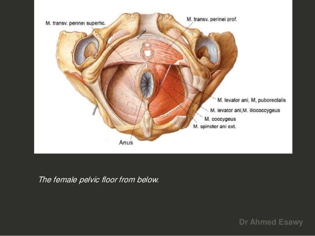 Anal Perianal Imaging Part 1 Ct Mri Anatomy Dr Ahmed Esawy
48 adductor longus muscle this muscle is the most anterior of the adductor group of muscles in the thigh.

Mri anatomy pelvis. Atlas of mri of the male pelvis. This webpage presents the anatomical structures found on hip mri. Magnetic resonance mr imaging is a valuable technique for the non invasive evaluation of the female pelvic region for example diagnosing or staging developmental anomalies leiomyomas adenomyosis vaginal neoplasms endometrial or cervical carcinoma.
This mri female pelvis sagittal cross sectional anatomy title tool is absolutely free to use. The muscle originates from the body of the pubis and attaches to the pectineal line and proximal part of the linea aspera of femur. Mri provides superior soft tissue contrast resolution for imaging the anatomy best seen in t1 weighted and pathology best seen on t2 weighted of the pelvis 3.
Knee shoulder shoulder arthrogram ankle elbow wrist hip contact. Mri of the pelvis may be more focused on the organs soft tissues and vessels rather than on the bones themselves. Mri of the male pelvis.
1 corpus cavernosum 2 corpus spongiosum bulb of the penis 3 ramus ischium 4 ischiocavernosus m. In many instances mri may be used to further clarify or confirm a diagnosis from another imaging modality. Click on a link to get t1 axial view t1 coronal view.
Mri of the pelvis is also beneficial for pre op planning and cancer staging. Mri of the hip. Stanford bone tumor bayesian network issssr msk lectures for residents ocad msk cases from around the world stanford msk mri atlas has served almost 800000 pages to users in over 100 countries.
Use the mouse scroll wheel to move the images up and down alternatively use the tiny arrows on both side of the image to move the images. 7 gluteus maximus m. Mri of the male pelvis.
This mri male pelvis axial cross sectional anatomy tool is absolutely free to use. It is enervated by the obturator nerve. Anatomy of the female pelvis mri atlas of the human body using cross sectional imaging.
5 anal canal 6 sphincter ani externus m. Use the mouse to scroll or the arrows.
 Getting Ready For An Mri Of Your Pelvis Sansum Clinic
 Download Ct And Mri Interactive Atlas Of Cross Sectional Anatomy 1 1e
 Ct Anatomy Of The Pelvis
 11 Axial Mri Of The Pelvic Structures Cg Coccygeus Muscle
 Musculoskeletal Mri
 Hip Mri

 Mri Pelvis Anatomy Free Male Pelvis Axial Anatomy
%20image%2018.jpg) Mri Female Pelvis Anatomy Free Mri Sagittal Cross
Mri Female Pelvis Anatomy Free Mri Sagittal Cross
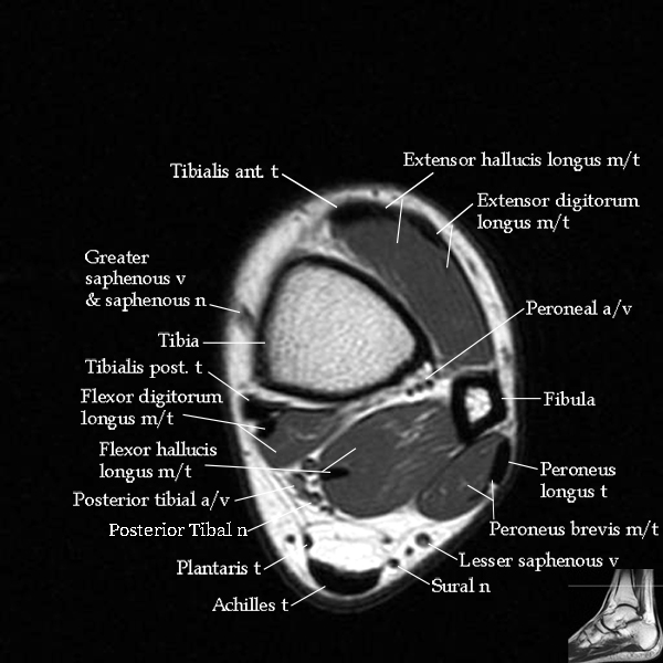 Mri Ankle Anatomy
 Normal Mri Hip Radiology Case Radiopaedia Org
 Pelvis And Perineum Radiology Key

Mri Pelvis Anatomy Free Male Pelvis Axial Anatomy
 Lumbar Spine Anatomy On Mri Magnetic Resonance Imaging
 Module 5 Pelvis Imaging
 Female Pelvis Anatomy Free Axial Cross Sectional Anatomy
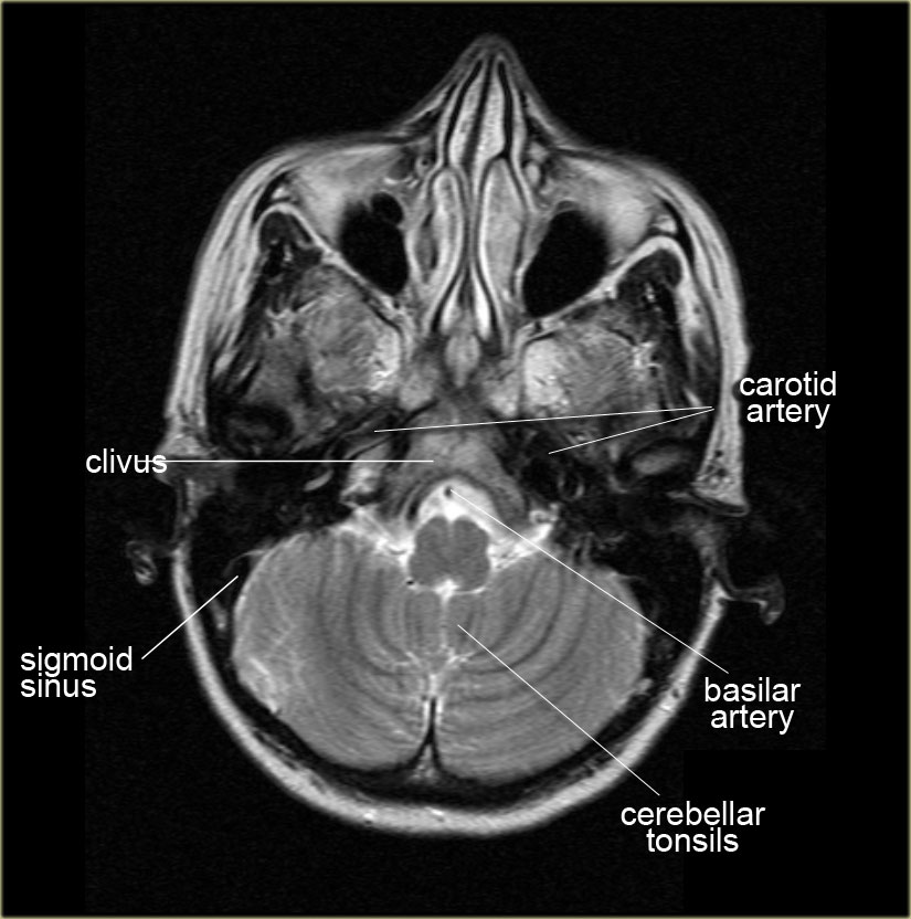 The Radiology Assistant Brain Anatomy


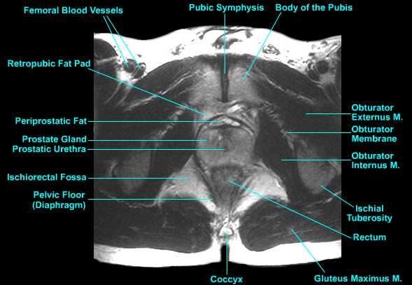
 The Male Pelvis Mr Anatomy Atlas Of The Prostate Bladder
 Sectional Anatomy Quiz 7 Pelvis Flashcards Quizlet

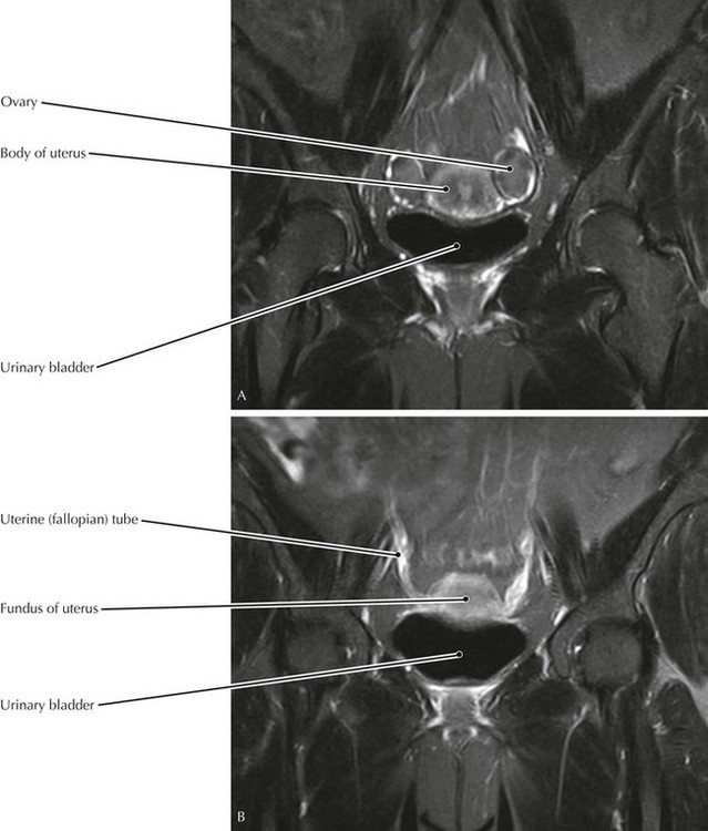
 Mri Of The Female Pelvis
 Chirogeek Com
 The 5 Things We Wish You Knew About Pudendal Neuralgia
 Normal And Variant Pelvic Anatomy On Mri Semantic Scholar
 Figure 1 From Does Bilateral Sacrospinous Fixation With


Posting Komentar
Posting Komentar