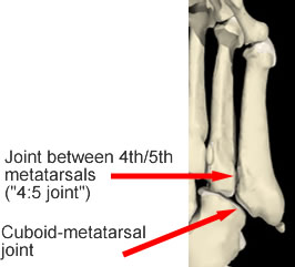
These bones do not have individual names rather these are numbered from inside to outside. The base articulates with the cuboid and with the fourth metatarsal.
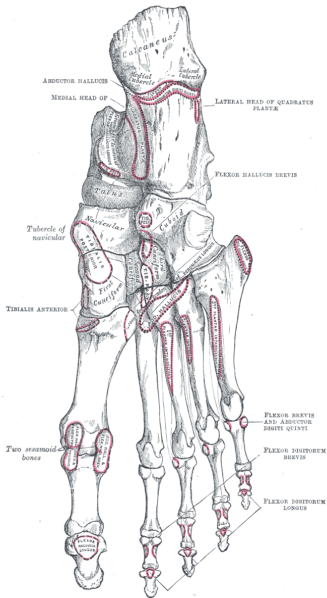
 File An Atlas Of Human Anatomy For Students And Physicians
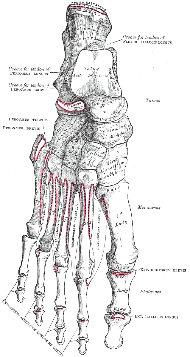
The second plantar interossei muscle originates from the medial side of the base and shaft.
Metatarsal anatomy. The first second third fourth and fifth metatarsal often depicted with roman numerals. The term describes pain and inflammation in the ball of the foot. The muscle attachments of the third metatarsal are the following.
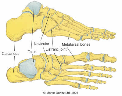
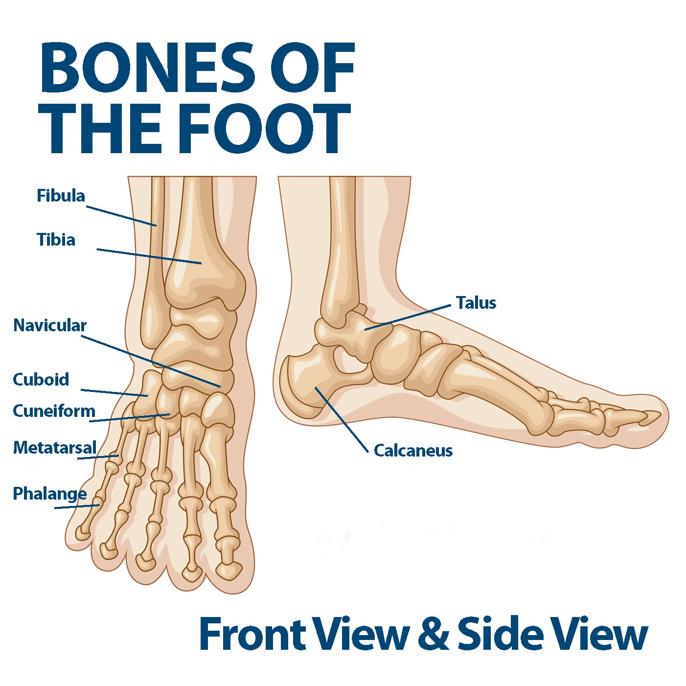
Each metatarsal has a similar structure. The metatarsals are the bones of the foot present between the heel tarsus of the foot and the toes. They work with connective tissues ligaments and tendons to provide movement in the foot.
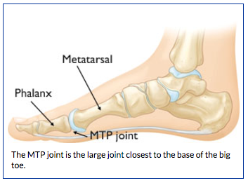
The metatarsal bones are connected to the bones of the toe or phalanges at the knuckle of the toe or metatarsophalangeal joint. Lateral shaft 3rd dorsal interosseus. The fifth metatarsal forms the mobile lateral foot border.
It is often thought of as a symptom of other conditions rather than as a specific disease. The metatarsal bones run from the tarsus forming the tarsometatarsal joints to the base of proximal phalanges forming the metatarsophalangeal joints. The horizontal head of the adductor hallucis also originates from the lateral side.
Fourth metatarsal bone the third and fourth dorsal interossei muscles originates from fourth metatarsal bone. They are convex dorsally and consist of a head neck shaft and base distal to proximal. Medial shaft 2nd dorsal interosseus and 1st plantar interosseus.
Metatarsals are convex in shape arch upward are long bones and give the foot its arch. 2nd 5th metatarsal have distal intermetatarsal ligaments that maintain length and alignment with isolated fractures implicated in formation of interdigital mortons neuromas multiple metatarsal fractures lose the stability of intermetatarsal ligaments leading to increased displacement. The anatomy of these joints and their ligaments has not been described in detail.
These are a group of five long cylindrical bones that are collectively called metatarsus. Metatarsalgia is a common overuse injury. Proximally tarsometatarsal joints between the metatarsal bases and the tarsal bones.
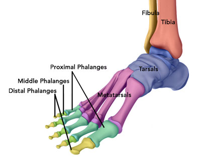
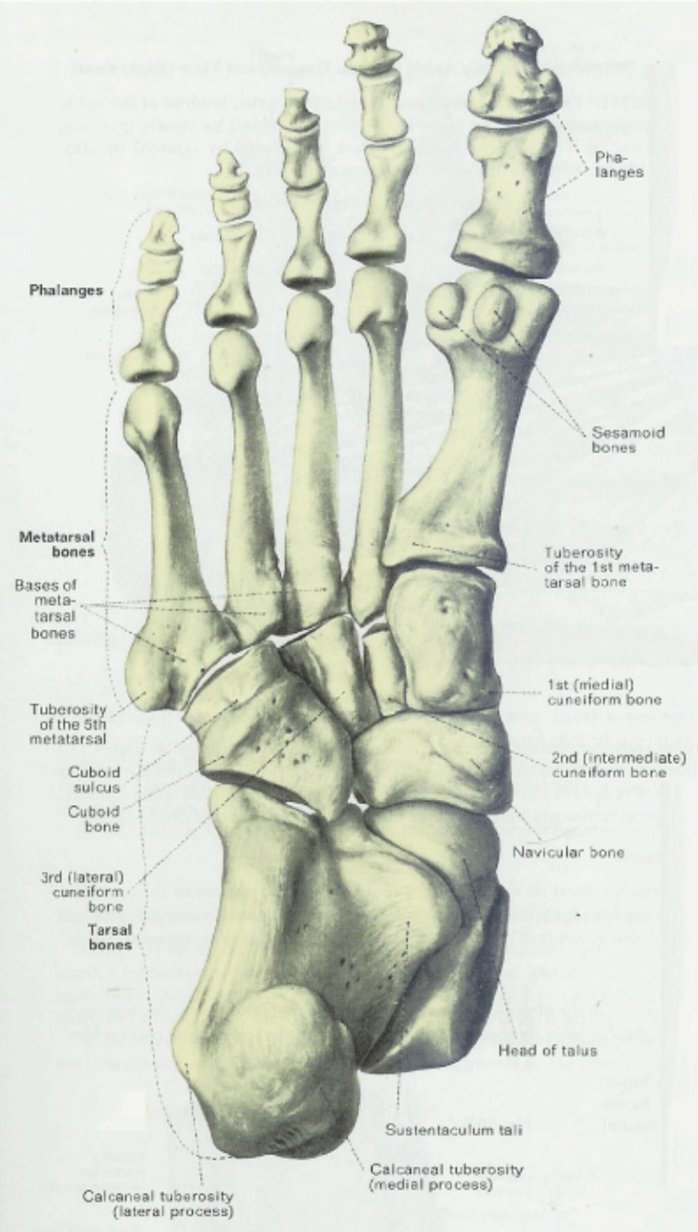
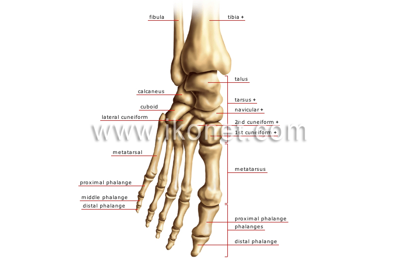
They have three or four articulations. Lacking individual names the metatarsal bones are numbered from the medial side the side of the great toe. The metatarsal bones or metatarsus are a group of five long bones in the foot located between the tarsal bones of the hind and mid foot and the phalanges of the toes.
It has a broad base expanded laterally to form the tuberosity a narrow shaft and a fairly small head.
 Metatarsal Anatomy
 Metatarsal Fracture Broken Foot In Depth Ankle Foot
 Ankle Injury Treatment Atlanta Ga Ankle Arthroscopy
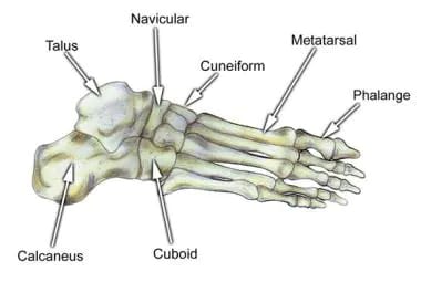
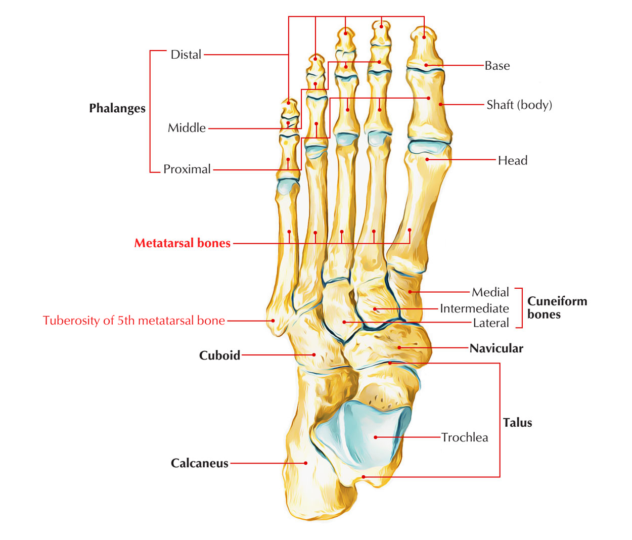
 Tarsal Metatarsal And Phalanges Of The Foot

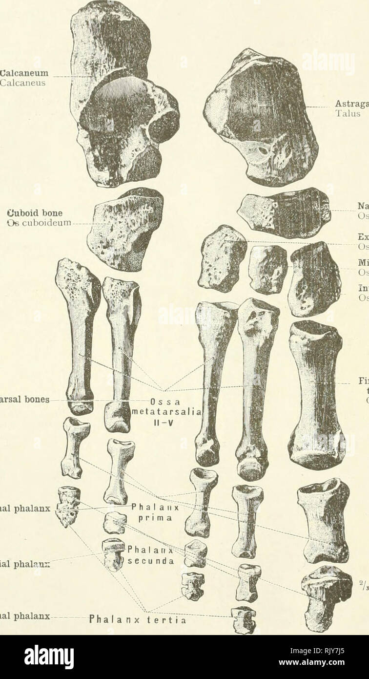 An Atlas Of Human Anatomy For Students And Physicians
 Metatarsals Approach Lateral Approach To The 5th
 Metatarsalgia Florida Orthopaedic Institute
 Foot Bone Anatomy Overview Tarsal Bones Gross Anatomy

 Broken Foot Pictures Symptoms Treatment Healing Time
Anatomy Of Turf Toe Bouldercentre For Orthopedics Spine
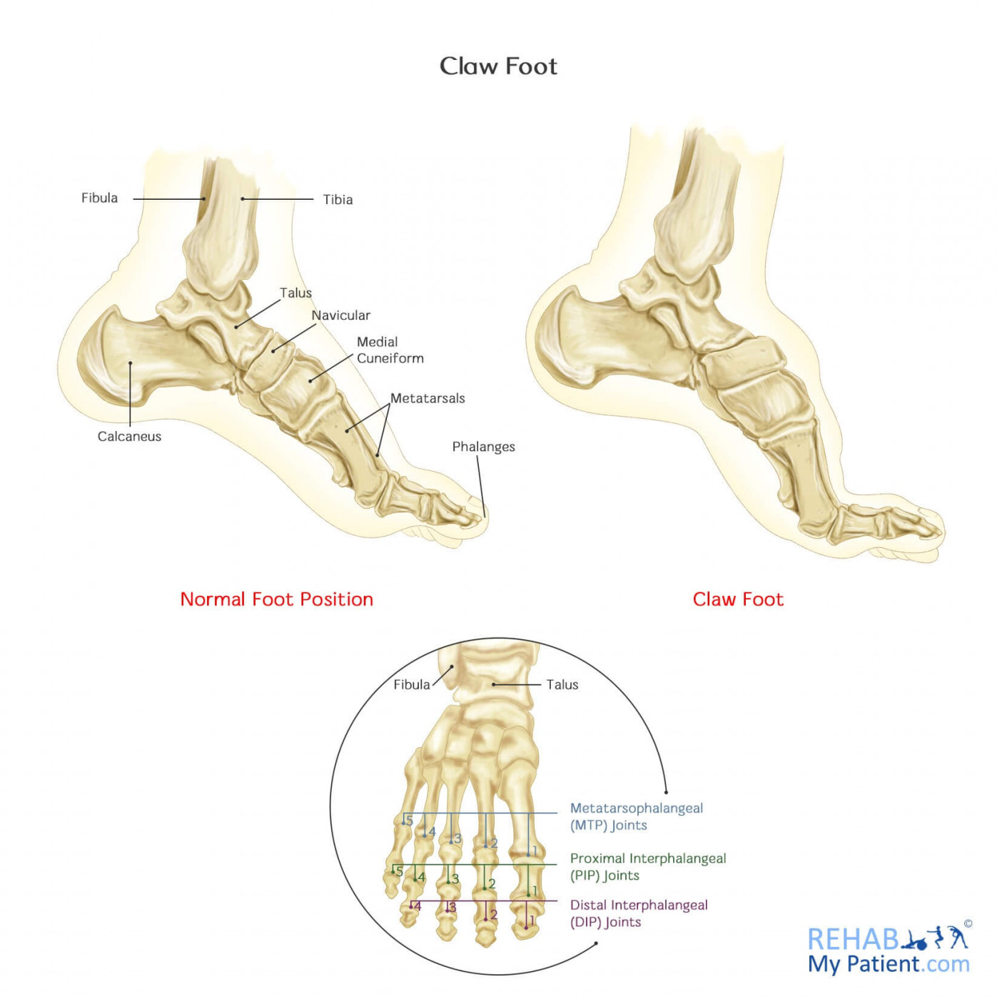 Claw Foot Rehab My Patient
 Metatarsals Approach Dorsal Intermetatarsal Approach
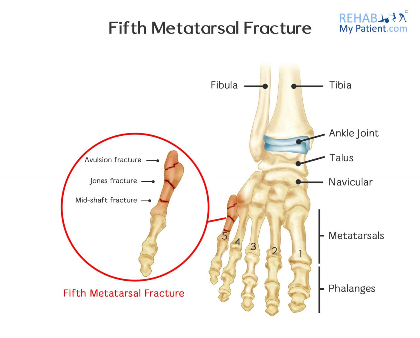 Fifth Metatarsal Fracture Rehab My Patient
Deep Transverse Metatarsal Ligament
 The Fifth Metatarsal Earth S Lab
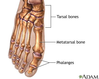 Bunion Removal Series Normal Anatomy Medlineplus Medical
 The Foot And Toes Essentials Of Athletic Injury Management
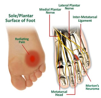 Morton S Neuroma Physiopedia
 Anatomy 101 Strengthen Your Big Toes To Build Stability
 5th Metatarsal Fractures
 Tarsal Metatarsal And Phalanges Of The Foot
 Metatarsal Bones Wikipedia
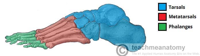 Bones Of The Foot Tarsals Metatarsals Phalanges
 Metatarsal Bone An Overview Sciencedirect Topics
 Anatomy Of Metatarsal Bones And Phalanges Bone And Spine
Plantar Metatarsal Arteries
 Second Metatarsal Stress Fractures Sciencedirect
 Ball Of Foot Pain Do The Bottoms Of Your Feet Toes Hurt
 Anatomy Of Metatarsal Bones And Phalanges Bone And Spine


Posting Komentar
Posting Komentar