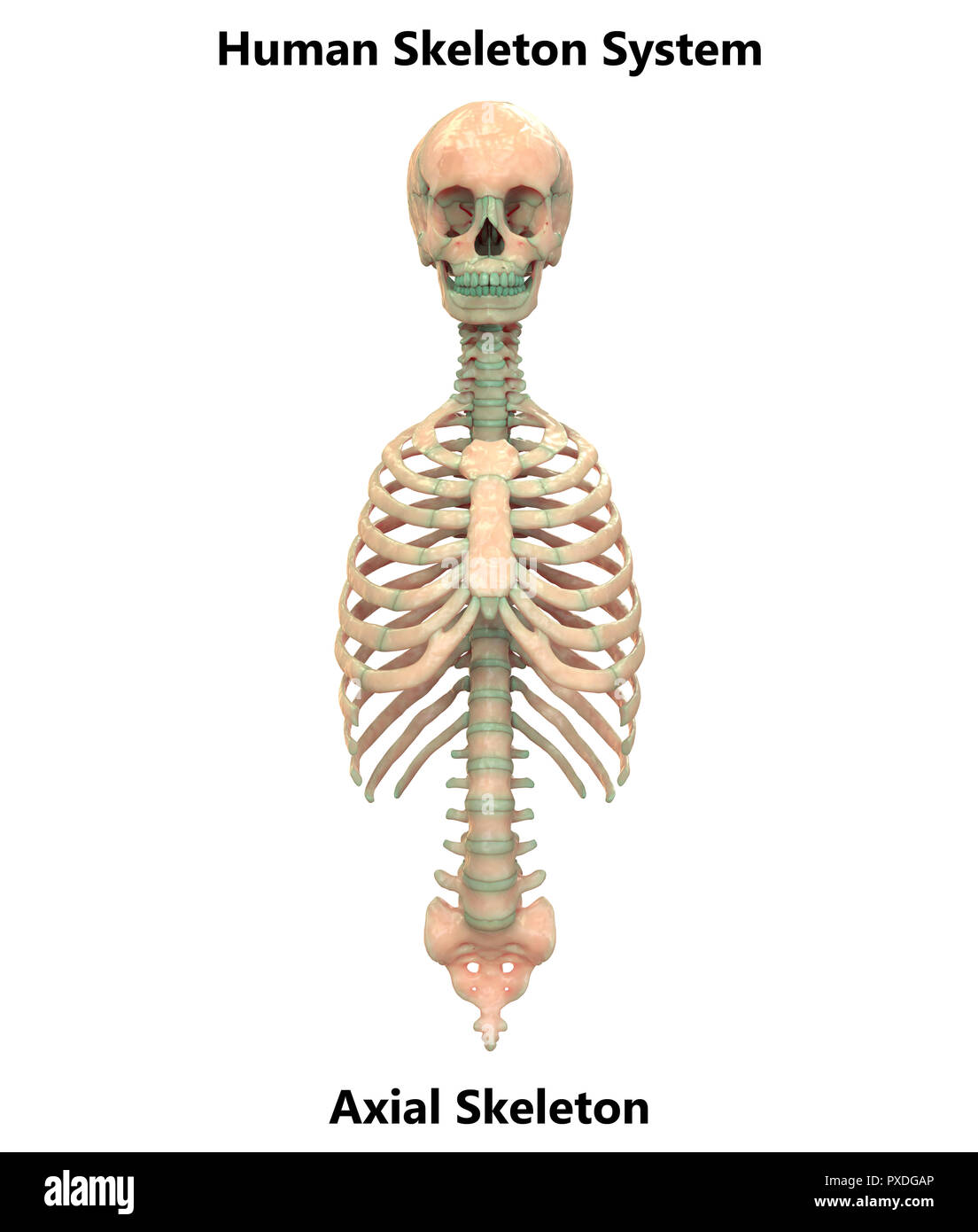
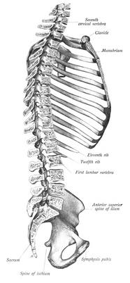
The rib cage is comprised of 12 pairs of ribs and the sternum. In radiology an axial image is one obtained by rotating around the axis of the.
 Human Skeleton System Appendicular And Axial Skeleton
The skull consists of the cranium and the facial bones.

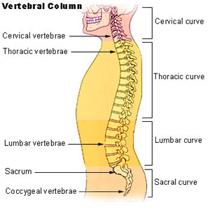
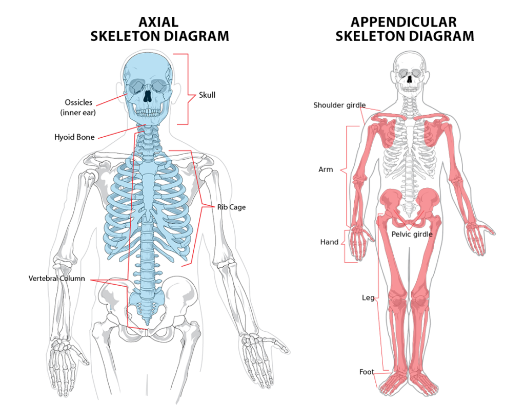
Axial anatomy. Mathematics a line ray or line segment with respect to which a figure or object is symmetrical. The skull consists of the cranial bones and the facial skeleton. In human skeletal system these are 1 the axial comprising the vertebral columnthe spineand much of the skull and 2 the appendicular to which the pelvic hip and pectoral shoulder girdles and the bones and cartilages of the limbs belong.
Relating to an axis. The bones of the appendicular skeleton the limbs and girdles append to the axial skeleton. Axis anatomy by the atlanto axial joint it forms the pivot upon which the first cervical vertebra the atlas which carries the head rotates.
This mri brain cross sectional anatomy tool is absolutely free to use. Axial skeleton skull bones. Must open the lower jaw of skull to identify this prominent foramen on the medial aspect of the mandibular ramus.
In humans it separates the superior from the inferior or put another way the head from the feet. A reference line from which distances or angles are measured in a coordinate system such as the x axis and y axis in the cartesian coordinate system. Skull bones protect the brain and form an entrance to the body.
In dentistry relating to or parallel with the long axis of a tooth. The most distinctive characteristic of this bone is the strong odontoid process known as the dens which rises perpendicularly from the upper surface of the body. Permits the passage of the nerve involved with tooth sensation and is the site where the dentist injects novacain to prevent pain while working on the lower teeth.
The axial skeleton includes the bones that form the skull laryngeal skeleton vertebral column and thoracic cage. A transverse also known as axial or horizontal plane is parallel to the ground. Human anatomy the following terms are defined in reference to the anatomical model being in the upright orientation standing.
Anatomy ct axial brain form no 18. At birth the majority have 32 34 separate. Relating to or situated in the central part of the body in the head.
There are 14 facial bones that form the lower part of the skull. The cranial bones compose the top and back of the skull and enclose the brain. Use the mouse scroll wheel to move the images up and down alternatively use the tiny arrows on both side of the image to move the images.
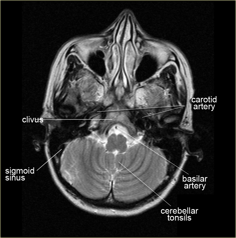
6 frontal bone 27 occipital bone 32 optic nerve 37 basilar artery 40 hemisphere of cerebellum 43 frontal sinus 45 sigmoid sinus 46 internal carotid artery 47 sphenoid bone 49 medulla oblongata 50 external auditory meatus 51 spinal central canal.
 Mri Anatomy Free Mri Axial Brain Anatomy
 Ct Cross Sectional Anatomy Abdomen Axial View
 Abdomen Anatomy Mri Abdomen Axial Anatomy Free Cross
 Axial Muscles Trunk Muscles Anatomy Support
 Seer Training Axial Skeleton 80 Bones
 Free Anatomy Quiz The Axial Skeleton Quiz 1
 Axial Skeleton Wikipedia

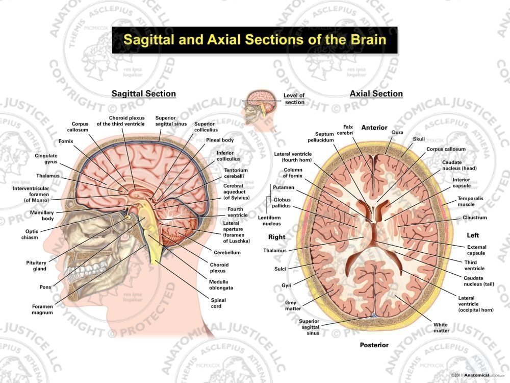 Sagittal And Axial Sections Of The Brain
 Axial Skeleton Human Anatomy Examville Com Free Study Aids
 Skeletal System Axial And Appendicular Skeletons Preview Human Anatomy Kenhub
 Anatomy Of Penis A And B Illustrations A Axial View B
 The Radiology Assistant Brain Anatomy
 Bear Gulch Fish Anatomy Axial And Caudal
 Appendicular Axial Skeleton Anatomy Anatomy Info
 Axial Brain Mri Anatomy
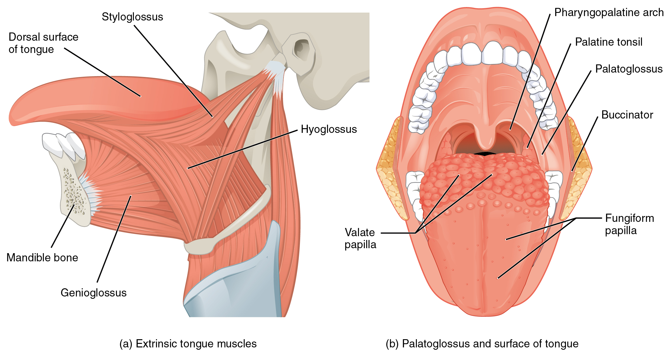 11 3 Axial Muscles Of The Head Neck And Back Anatomy And
 Anatomy Axial Skeleton Test Pt 1 Diagram Quizlet
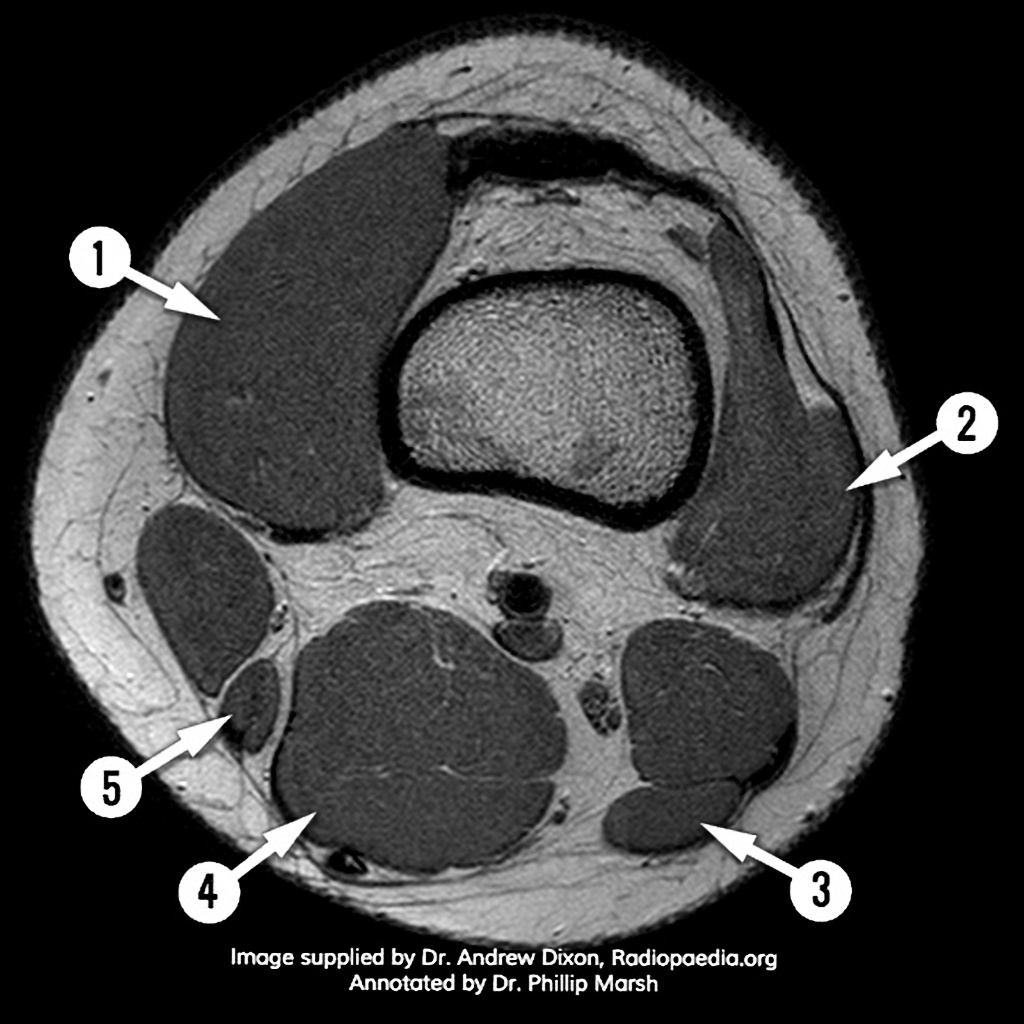 Mri Knee Axial Anatomy Quiz Radiology Case
 Mri Neck Anatomy Free Mri Axial Neck Cross Sectional Anatomy
 Overview Of Skull Base Anatomy
Mri Anatomy Free Mri Axial Brain Anatomy
 Radiology Basics Chest Anatomy
 Ct Neck Axial Anatomy Radiologypics Com
 Axial Skeleton Diagram Skeleton Anatomy Axial Skeleton
 Cross Sectional Anatomy Of The Brain
 Ct Chest Anatomy Axial Anatomy Of The Thorax Studykorner
 Mri Anatomy Free Mri Axial Brain Anatomy


Posting Komentar
Posting Komentar