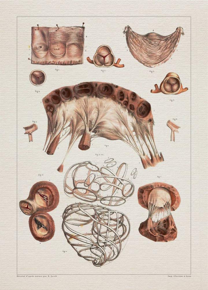
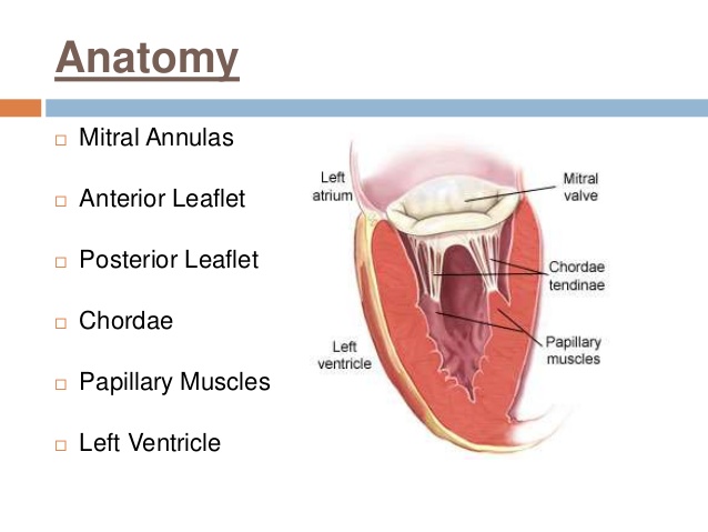
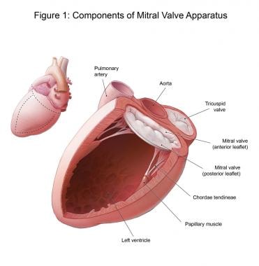
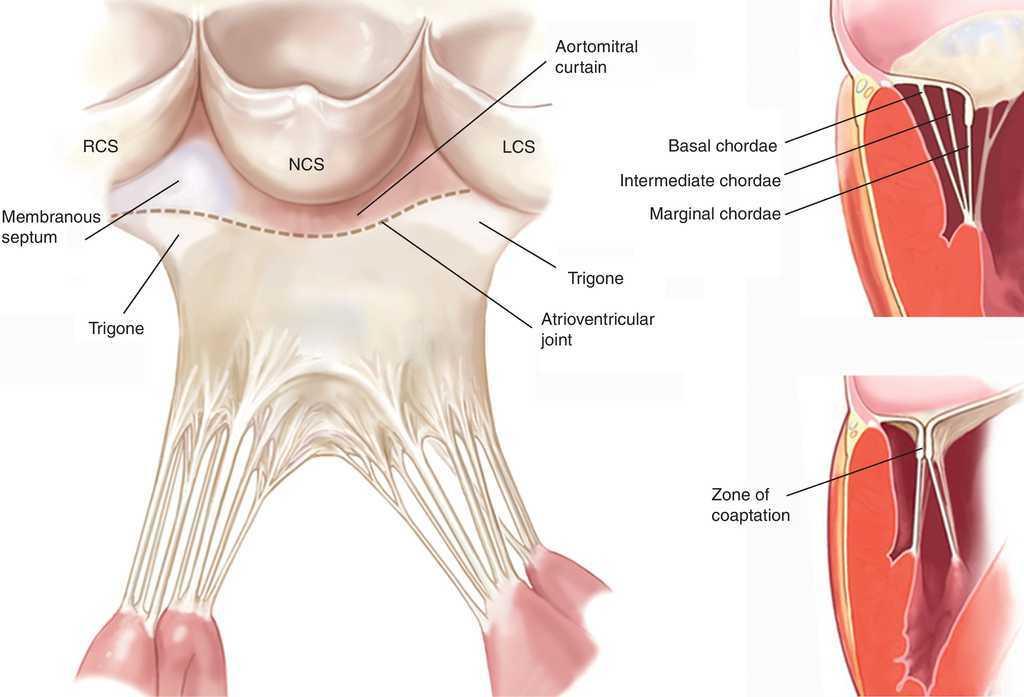
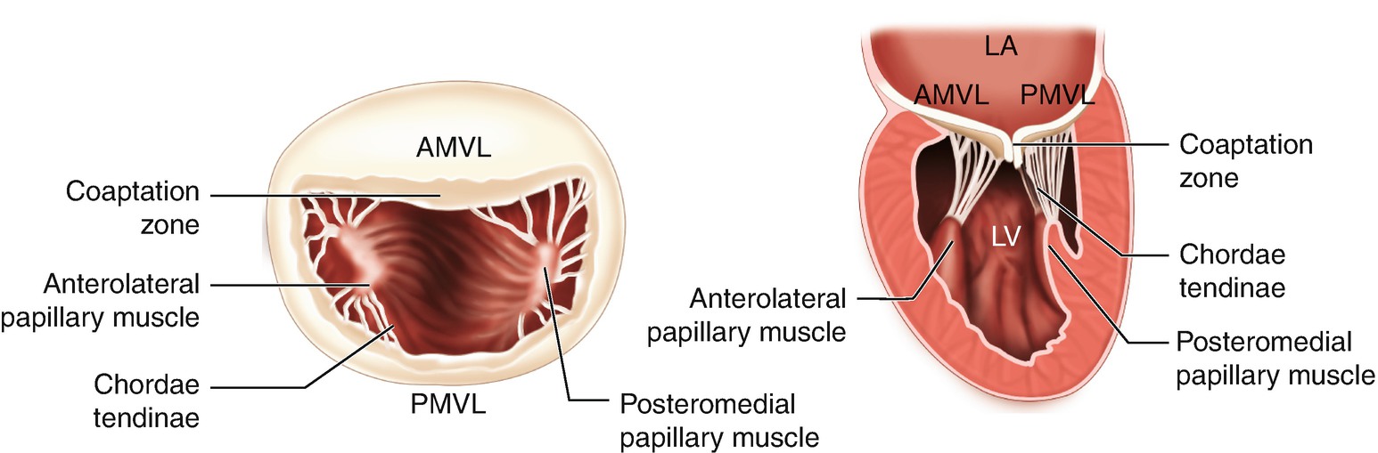
Anatomy of the mitral valve. The chordae tendinae are fan shaped connective.
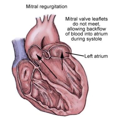
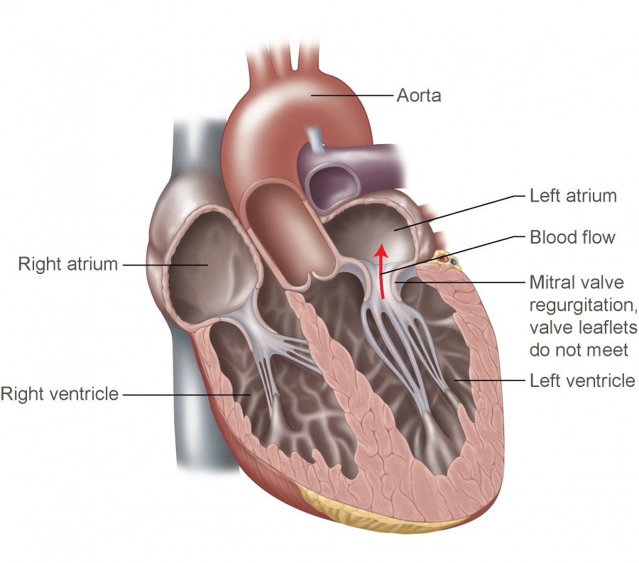
 Mitral Regurgitation
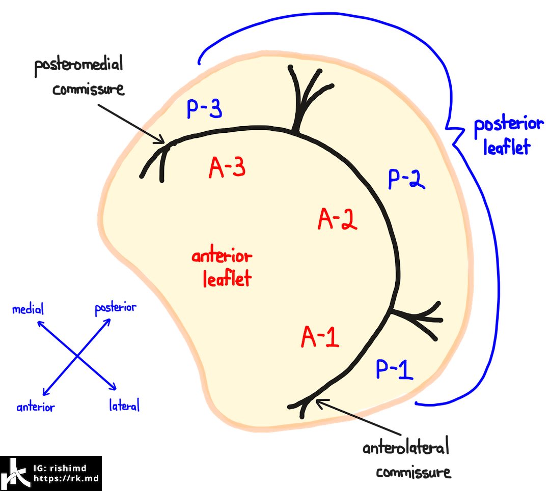
The mitral valve has two leaflets.
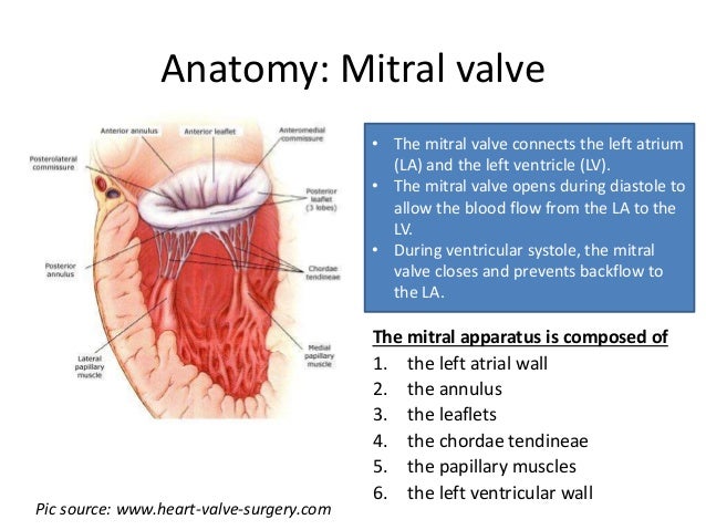
Anatomy of mitral valve. Thus it is in close proximity to the aortic valve left and acoronary cusp. The mitral valve also known as the left atrioventricular valve or bicuspid valve connects the left atrium with and the left ventricle. The mitral valve is a complicated 3d structure composed of multiple distinct anatomical components.
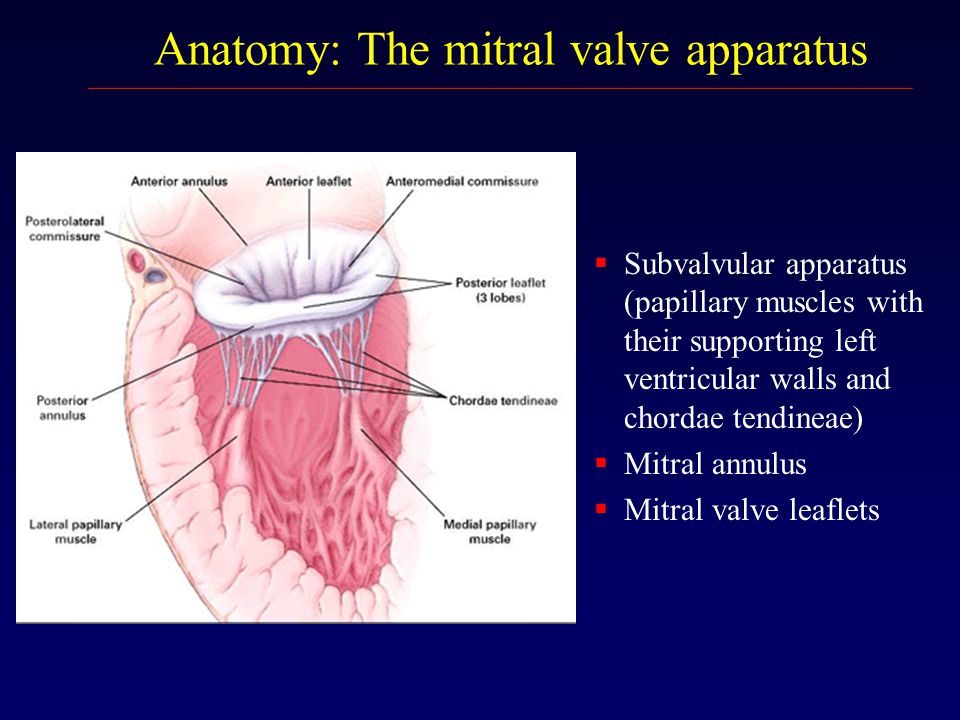
Optimal interaction of these different elements comprising the annulus commissures leaflets chordae tendinae papillary muscles and left ventricle is crucial for its functional integrity. Note the wider middle scallop. Thin and pliable leaflets that contain scallops which represent segmental markers.
These are projections that open and close. Five mitral valve components 1. The mitral apparatus has very specific details that make.
What do the mitral valves different parts do. Imaging the mitral valve mv requires an understanding of the normal anatomy and how this complex structure is altered by disease states. The mitral valve opens during diastole to allow the blood.
The free edge of the mural leaflet has two clefts red arrows giving this leaflet the appearance of three scallops. The posterior leaflet is divided into scallops the anterior leaflet is longer and is positioned adjacent to the intervalvular fibrosa. The normal mitral valve apparatus is a dynamic three dimensional system that allows brisk left ventricular lv blood inflow during diastole and ensures unidirectional heart pump function by sealing the left atrium from the lv during systole.
Mitral leaflets scallops. The mv is composed of several structures working in synchrony to open during diastole and close in systole effectively within the high pressure systemic environment. Guarding the inlet to the left ventricle the mitral valve prevents backflow to the left atrium during ventricular systole.
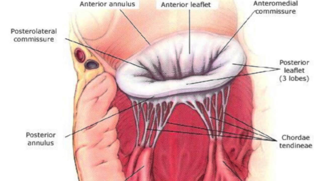
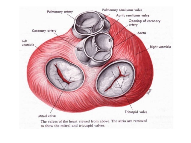
The commissures of the mitral valve are the areas where the anterior. Mitral valve according to walmsley 1 it was andreas vesalius who suggested the picturesque term mitral to describe the left atrioventricular valve owing to its resemblance to a plan view of the bishops mitre. A the atrial aspect of the mitral valve shows the arrangement of the two leaflets between the commissures.
 Anatomy Of The Mitral Valve Apparatus A Mitral Valve And
 Amazon Com Anatomy Mitral Valve Heart Print Sra3 12x18

 Percutaneous Mitral Valve Repair Background Indications
 Mitral Valve Regurgitation Temple Health
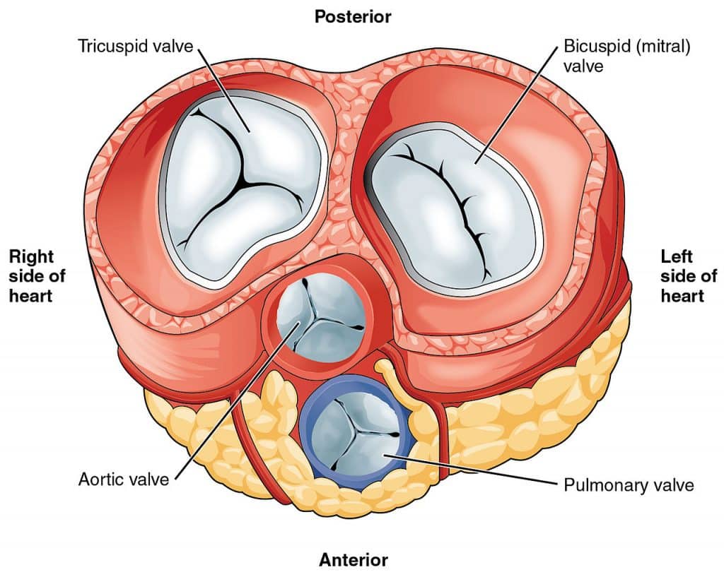
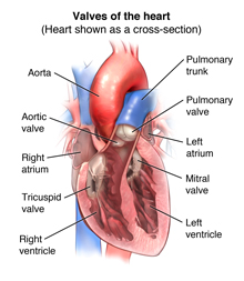 Anatomy And Function Of The Heart Valves
 Mitral Valve Anatomy Overview Gross Anatomy Microscopic
 Endoscopic Robotic Mitral Valve Repair
 The Anatomy Of Native Mitral Valve Apparatus Medscape 2017
:max_bytes(150000):strip_icc()/human-heart-circulatory-system-598167278-5c48d4d2c9e77c0001a577d4.jpg) Av And Semilunar Heart Valves
Av And Semilunar Heart Valves
 Science Source Heart Anatomy Illustration
 Mitral Valve Anatomy Ppt By Kunwar Sidharth
 The Heart Valves Tricuspid Aortic Mitral Pulmonary
 Mitral Regurgitation Francesca N Delling Md July 8 Ppt
 Left Ventricle An Overview Sciencedirect Topics

 Surgical Echocardiography Of The Mitral Valve Revista
 Mitral Valve Anatomy Echocardiography And Surgical
 Figure 1 From The Left Ventricle In Dogs With Myxomatous
 Mitral Valve Regurgitation Latest News Cardiomyopathy Uk
 Mitral Valve Anatomy Britannica
 Heart Valve Anatomy Mitral Valve Heart Png Clipart Free
 Mitral Valve Anatomy Rk Md

 Mitral Valve Anatomy Source 24 Download Scientific
 Anatomy And Physiology Of The Mitral Valve Springerlink
Chapter 1 Surgical Anatomy Of The Aortic And Mitral Valves
Chapter 1 Surgical Anatomy Of The Aortic And Mitral Valves
Systolic Anterior Motion Of The Mitral Valve Chordae With Dr David Adams


Posting Komentar
Posting Komentar