There are also rotational movements at the knee joint. Three bones meet to form your knee joint.
 Knee Leg Atlas Of Anatomy
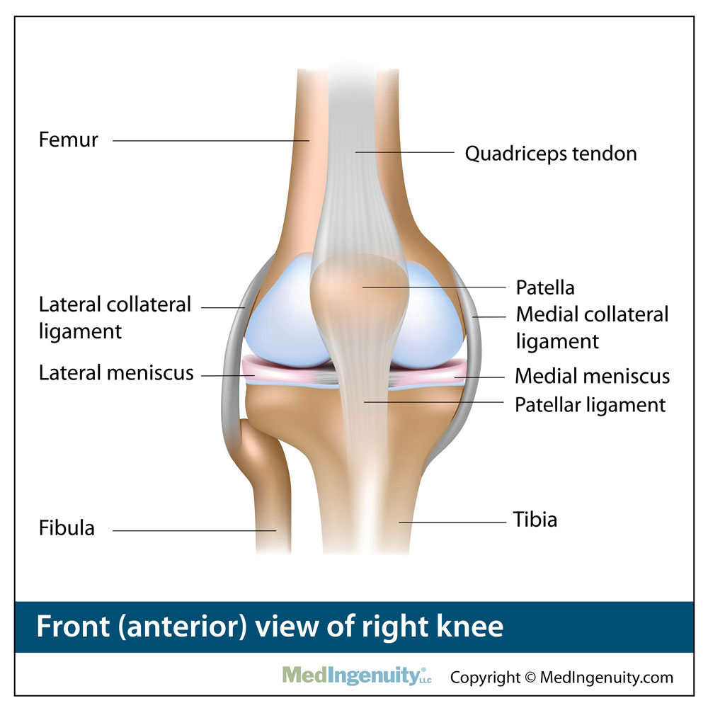
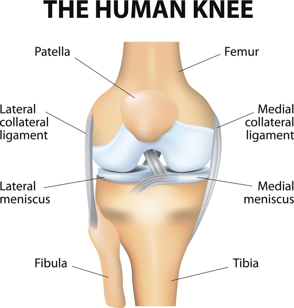
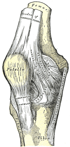
Anteromedial aspect of right knee the ligaments surrounding the knee joint offer stability by limiting movements and together with the menisci and several bursae protect the articular capsule.
Knee anatomy right. In knee joint anatomy they are the main stabilising structures of the knee acl pcl mcl and lcl preventing excessive movements and instability. The knee is a hinge joint that is responsible for weight bearing and movement. 1 the tibiofemoral joint where the tibia meet the femur 2 the patellofemoral joint where the kneecap or patella meets the femur.
Bones cartilage ligaments and tendons. The most common ligament injuries are acl tears mcl tears pcl tears and knee sprains which occur when the ligaments are overstretched. Use the mouse to scroll.
Knee anatomy function and common problems the knee joint is a synovial joint which connects the femur thigh bone the longest bone in the body to the tibia shin bone. The knee is a complex joint that flexes extends and twists slightly from side to side. There are two main joints in the knee.
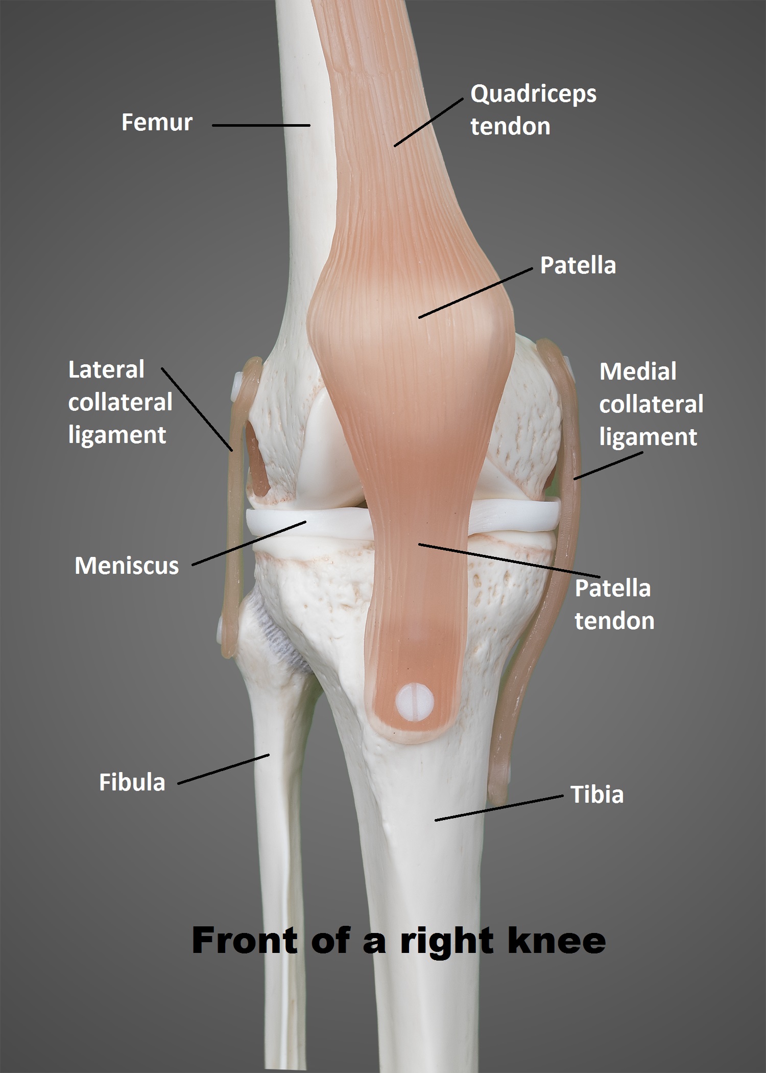
The knee joins the thigh bone femur to the shin bone tibia. The knee is one of the largest and most complex joints in the body. The primary function of the knee is a hinged of the lower extremity.
Your thighbone femur shinbone tibia and kneecap patella. It is made up of four main things. The smaller bone that runs alongside the tibia fibula and the.
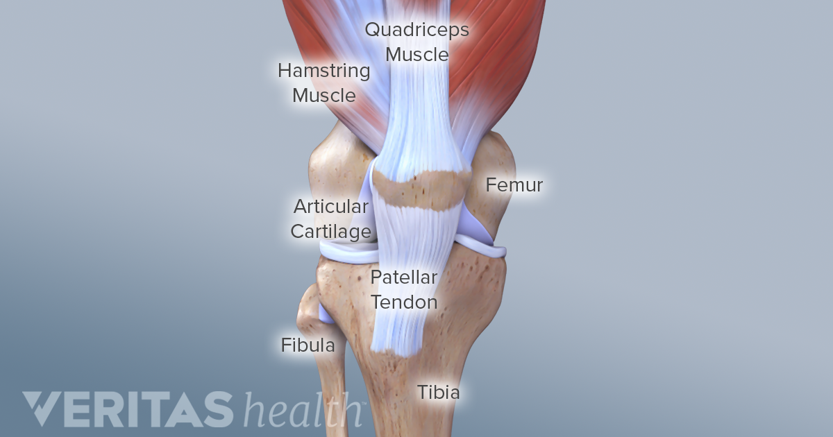
Knee function is determined in large part by the anatomy of the joint. The knee is the meeting point of the femur thigh bone in the upper leg and the tibia shinbone in the lower leg. Knee anatomy share on pinterest the knee is the most complex joint in the human body.
However the knee does not only been back and forth. The two menisci of the knee joint are pads of tissue which serve to limit friction in the knee. The fibula calf bone the other bone in the lower leg is connected to the joint but is not directly affected by the hinge joint action.
Intracapsular edit. The knee is the largest joint in the body and one of the most easily injured. The meniscus is a tissue made of cartilage that act as shock absorbers between the femur and tibia.
 Leg Anatomy Britannica
 Right Lateral Knee Anatomy
 Knee Joint Anatomy Medical Illustration Medivisuals
 14102 04b Tendons And Ligaments Of The Right Knee Anatomy
The Knee Ut Health San Antonio
 Anatomy Library Fort Worth Bone Joint Clinic
/188058334-crop-56aae7425f9b58b7d0091480.jpg) What Is Causing Your Knee Pain
What Is Causing Your Knee Pain
 Knee Surgery Knee Anatomy
 The Knee Anatomy Injuries Treatment And Rehabilitation
 Knee Anatomy
 Osteotomy Of The Knee Jonathan Frank Md Orthopedic
 Free Art Print Of Posterior View Of The Right Knee
 Inner Knee Pain Lotus Not Blooming Yoga Anatomy
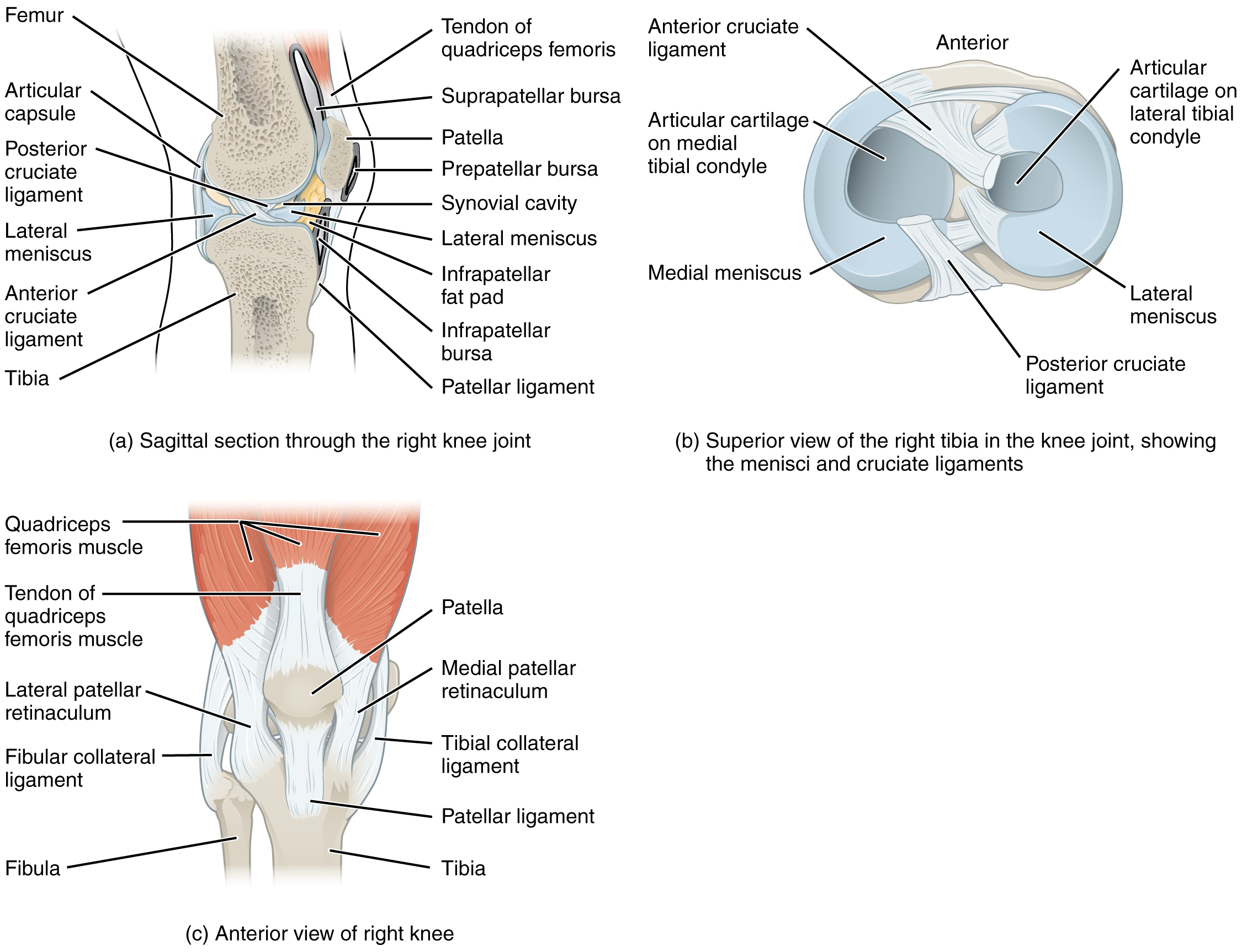 9 6 Anatomy Of Selected Synovial Joints Anatomy And Physiology
 The Knee Joint Human Anatomy
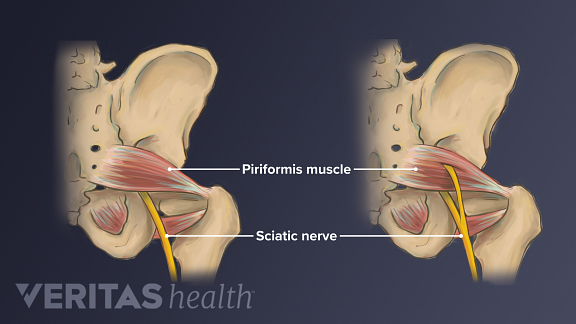 Sciatic Nerve Anatomy
 Ucsd S Practical Guide To Clinical Medicine
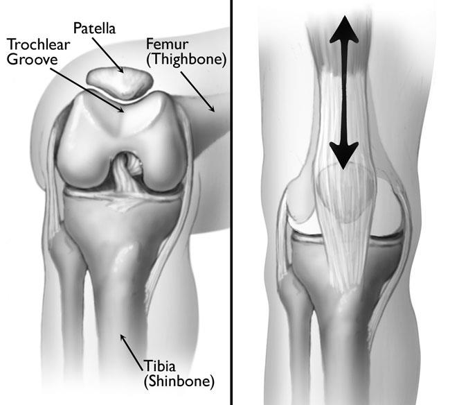 Patellofemoral Pain Syndrome Orthoinfo Aaos
 Right Above The Knee Amputation Ami 2018 Meeting
Search Anatomy Of The Knee Distracted
 Torn Meniscus Doctor Stock
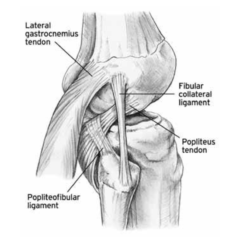 The Knee Resource Posterolateral Corner Injury
 Get Knee Deep In Knee Knowledge Yoga For Knee Rehab And
 Acl Solutions Acl Knee Anatomy And Diagram Images
 Amicus Injury Knee Prosthesis Lateral Tracking Patella
Stock Image Illustration Of Normal Knee Anatomy Top Left
 Articular Capsule Of The Knee Joint Wikipedia

 Muscles Of The Knee Anatomy Pictures And Information
 Knee Dislocations Everything You Need To Know Dr Nabil Ebraheim


Posting Komentar
Posting Komentar