However the muscular layers vary according to the region of the chest wall. Surgical anatomy of the chest wall.
 Chest Wall Tumors The Patient Guide To Heart Lung And
Thomas r gest phd more.
Anatomy of chest wall. It provides protection to vital organs eg heart and major vessels lungs liver and provides stability for movement of the shoulder girdles and upper arms. The chest wall protects the heart lungs and liver provides a flexible skeletal framework to stabilize the actions of the shoulder and arm and promotes respiratory movement all while reliably delivering more than 20000 breaths a day. This thoracic and pulmonary anatomy tool is especially designed for students of anatomy medical and paramedical studies.
This e anatomy module presents an illustrated anatomy of the lungs trachea bronchi pleural cavity and pulmonary vessels. This relation with the bone explains the risk for vessel wound in rib fractures. The chest wall is comprised of skin fat muscles and the thoracic skeleton.
Therefore in the dorsal and lateral chest the surgeon must proceed along the superior margin of the lower rib when puncturing in order to aspirate or to perform surgical procedures. Applied anatomy of the chest wall and mediastinum. The first rib is a short flat rib that is much wider and more curved than those previously described.
This mri chest thorax axial cross sectional anatomy tool is absolutely free to use. Use the mouse scroll wheel to move the images up and down alternatively use the tiny arrows on both side of the image to move the images. Navid pourtaheri md phd ms.
Anatomy of the chest and the lungs. Anatomy of the thoracic wall. Abstract the thoracic wall consists of the osseo cartilaginous throacic cage the interconnecting muscles the muscles on top the fascia the nerves and vasculature the subcutaneous tissue the skin and the mammary glands that lie within the subcutaneous tissue.
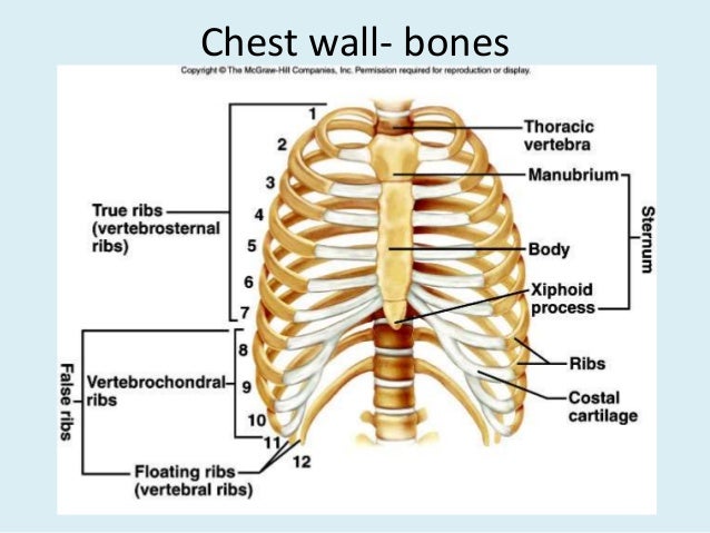
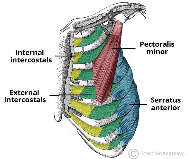
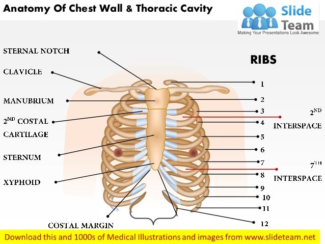
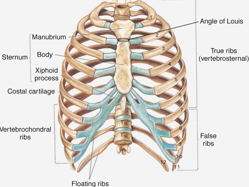
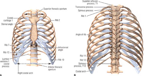
The spaces between the ribs are filled by the intercostal musculature which consists of three layers. Chest wall anatomy the chest is considered to be the area between the neck and the abdomen and contains many major organs as well as muscle groups. The skeleton of the thoracic wall is formed by the twelve thoracic vertebra posteriorly the sternum anteriorly and on each side by the twelve ribs and the respective costal cartilage.
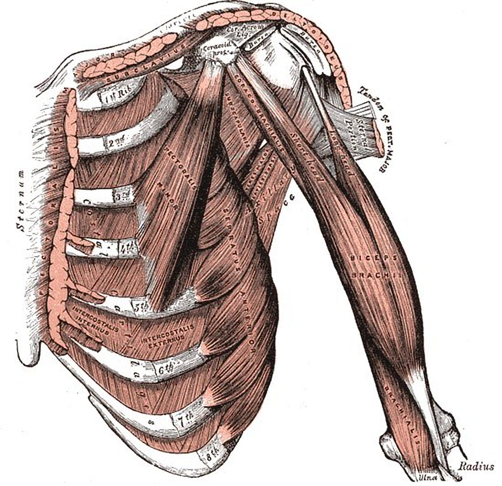
Anterior chest wall showing muscular attachments and neurovascular structures ribs 3 through 9 are typical ribs as described earlier while ribs 1 2 10 11 and 12 are atypical. The chest wall has 10 layers namely skin superficial fascia deep fascia serratus anterior layer for ribs containing intercostal muscles and endothoracic fascia from superficial to deep. For example they may include muscles like pectoralis major or latissimus dorsi.
 Anatomy Of Thorax 2
 Breast Anatomy Breast360 Org The American Society Of
 Thoracic Muscles Attachments Actions Teachmeanatomy
 Chest Wall Radiology Key
 0514 Anatomy Of Chest Wall And Thoracic Cavity Medical
 Chapter 18 Thoracic Wall Pleura Mediastinum Lung
 0514 Anatomy Of Chest Wall And Thoracic Cavity Medical
 Anatomy Of Chest Wall And Thoracic Cavity Medical Images For
 Muscles Of Anterolateral Chest Wall And Shoulder Radiology
 Thoracic Wall And Breast Illustrations
 Vintage 1950 S Frohse Chest Abdomen Viscera Human Anatomy Wall Chart
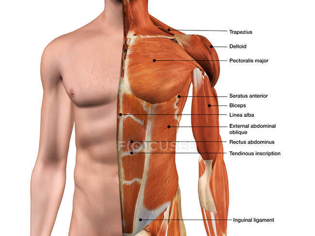 Male Anterior Thoracic Wall Chest Muscles Labeled On White
 The Muscles Connecting The Upper Extremity To The Anterior
 Chest Wall And Lung Anatomy And Physiology Ppt Download
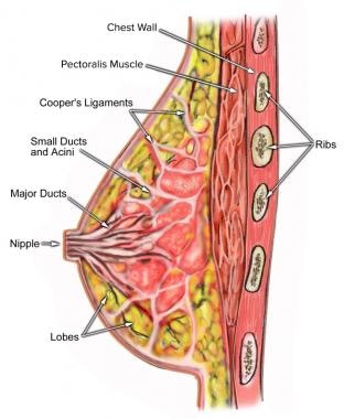 Breast Anatomy Overview Vascular Anatomy And Innervation
 Thorax Surface Anatomy 4 Edition
 Anatomy Of The Thoracic Wall Pulmonary Cavities And
 Applied Anatomy Of The Chest Wall And Mediastinum
 Anatomy Of Chest Wall Youtube
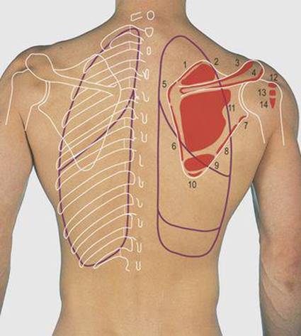
 Posterior Thoracic Wall 119 Download Scientific Diagram
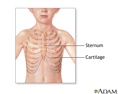 Pectus Excavatum Repair Series Normal Anatomy Medlineplus
 Figure 4 From Relevant Surgical Anatomy Of The Chest Wall
 Thoracic Wall And Breast Illustrations
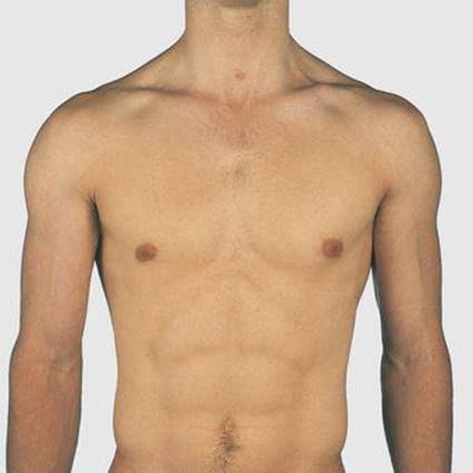 Thorax Surface Anatomy 4 Edition
 Surgical Anatomy Of The Chest Wall Springerlink
 The Pleurae Human Anatomy
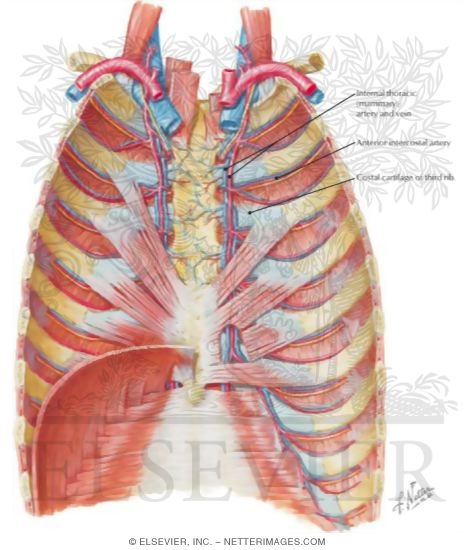 Anterior Thoracic Wall Anterior Thoracic Wall Internal View


Posting Komentar
Posting Komentar