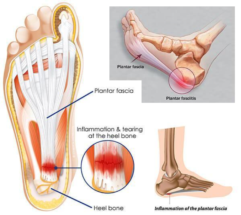
Diseases of the horses foot harry caulton reeks. Plantar fasciitis is the pain caused by degenerative irritation at the insertion of the plantar fascia on the medial process of the calcaneal tuberosity.
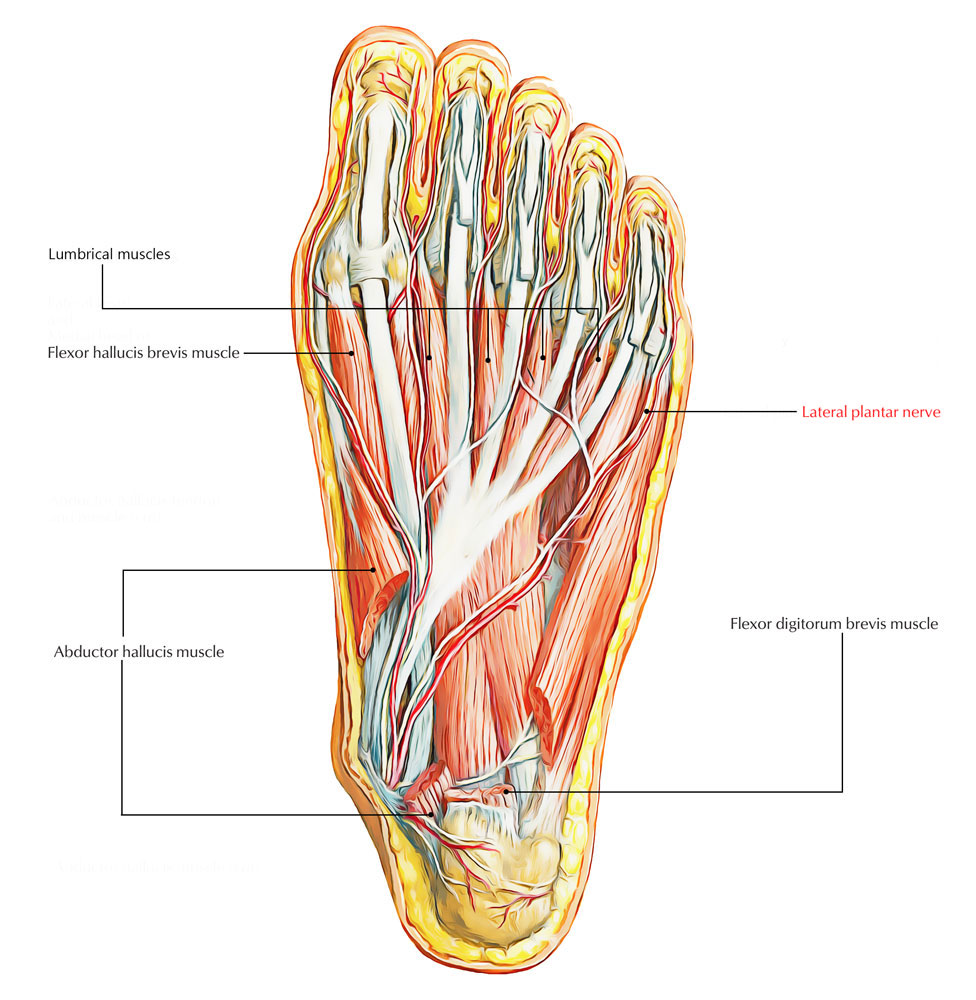 Easy Notes On Lateral Plantar Nerve Learn In Just 3
Or it may be of course that it was in the plantar aponeurosis the disease commenced.
Plantar anatomy. Plantar fasciitis is inflammation of the thick band of tissue also called a fascia at the bottom of your foot that runs from your heel to your toes. Running along the plantar groove it gains the plantar foramen. Arising predominantly from the calcaneal tuberosity the plantar fascia attaches distally through several slips.
It sits in the centre of the sole sandwiched between the plantar aponeurosis and the tendons of flexor digitorum longus. If talking about the skull the dorsal side is the top. These two terms used in anatomy and embryology refer to back dorsal and front or belly ventral of an organism.
In your case i suspect the cause is plantar fasciitis. Towards the front of the foot at the mid metatarsal level it divides into five sections each extending into a toe and straddling the flexor tendons. The plantar aponeurosis also known as the plantar fascia is a strong layer of white fibrous tissue located beneath the skin on the sole of the foot.
The dorsal from latin dorsum meaning back surface of an organism refers to the back or upper side of an organism. The pain may be substantial resulting in the alteration of daily activities. Anatomy of the plantar fascia.
Doctors once thought bony growths called heel. It attaches to the middle phalanges of the lateral four digits. Diseases of the horses foot harry caulton reeks.
Anatomy of the plantar fascia the plantar fascia is a complex structure that extends from the medial calcaneal tubercle the heel bone to the proximal phalanges of the toes the bone at the base of the toe at the metatarsophalangeal mtp joints. The strongest ligament is the plantar fascia which attaches the heel to the toes and helps to balance various parts of the foot as you walk. This image shows the anatomy of the plantar foot and is labeled with corresponding identification tags.
Originates from the medial tubercle of the calcaneus and the plantar aponeurosis. The plantar fascia or plantar aponeurosis forms part of the deep fascia of the sole of the foot and provides a strong mechanical linkage between the calcaneus and the toes. This median ridge fits into the cleft of the plantar cushion.
 Plantar Fasciitis Anatomy Body Disease En Fasciitis
 Ledderhose Disease Physiopedia
 Lateral Plantar Nerve Injury Following Steroid Injection For
 Layers Of The Plantar Foot Foot Ankle Orthobullets
 3cb Performance How Do You Know If You Have Plantar
 Select Chiropractic And Wellness Plantar Fasciitis
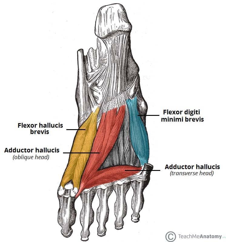
 Boards By The Numbers Anatomy Intrinsic Plantar Muscles Of
 Plantar Fasciitis Treatment Franciscan Health
 Plantar Fasciitis
 What Is Plantar Fasciitis Heelhealth
 Plantar Anatomy Exhibits
 Stock Illustration
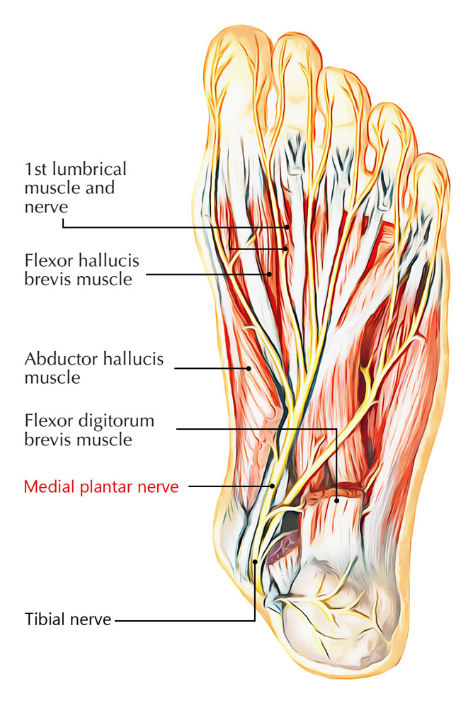 Easy Notes On Medial Plantar Nerve Learn In Just 4
 Plantar Fasciitis Fleet Feet Columbus
 Anatomy Of The Plantar Fascia Notes Adapted From
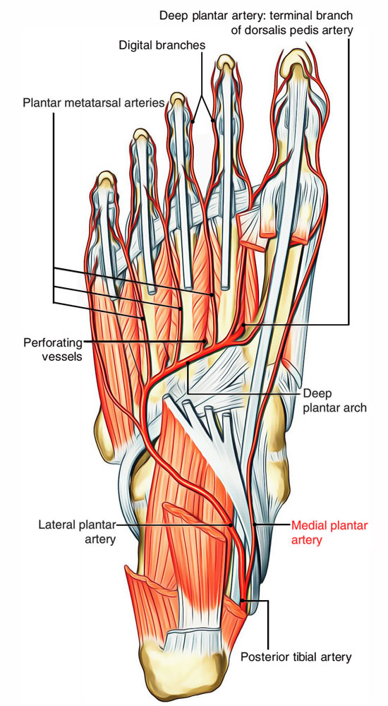 Arteries Of Foot Earth S Lab
![]() Central Plantar Muscles Of The Foot Anatomy Kenhub
Central Plantar Muscles Of The Foot Anatomy Kenhub
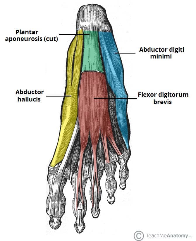 Muscles Of The Foot Dorsal Plantar Teachmeanatomy
 Layers Of The Plantar Foot Foot Ankle Orthobullets
 Ace Prosource April 2014 Understanding And Alleviating
 Muscles Of The Foot Dorsal Plantar Teachmeanatomy
 Plantar Fascitis Pinnacle Orthopaedics
 Plantar Fascia Anatomy Plantar Aponeurosis Anatomy


Posting Komentar
Posting Komentar