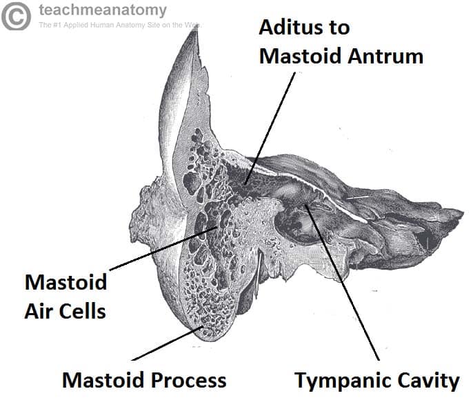
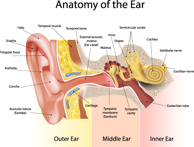
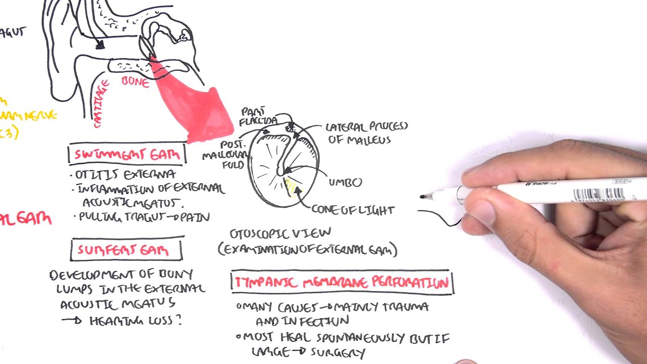
The eustachian tube helps to equalize the pressure in. The inner ear includes.

The area is pressurized.
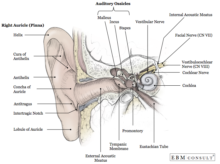
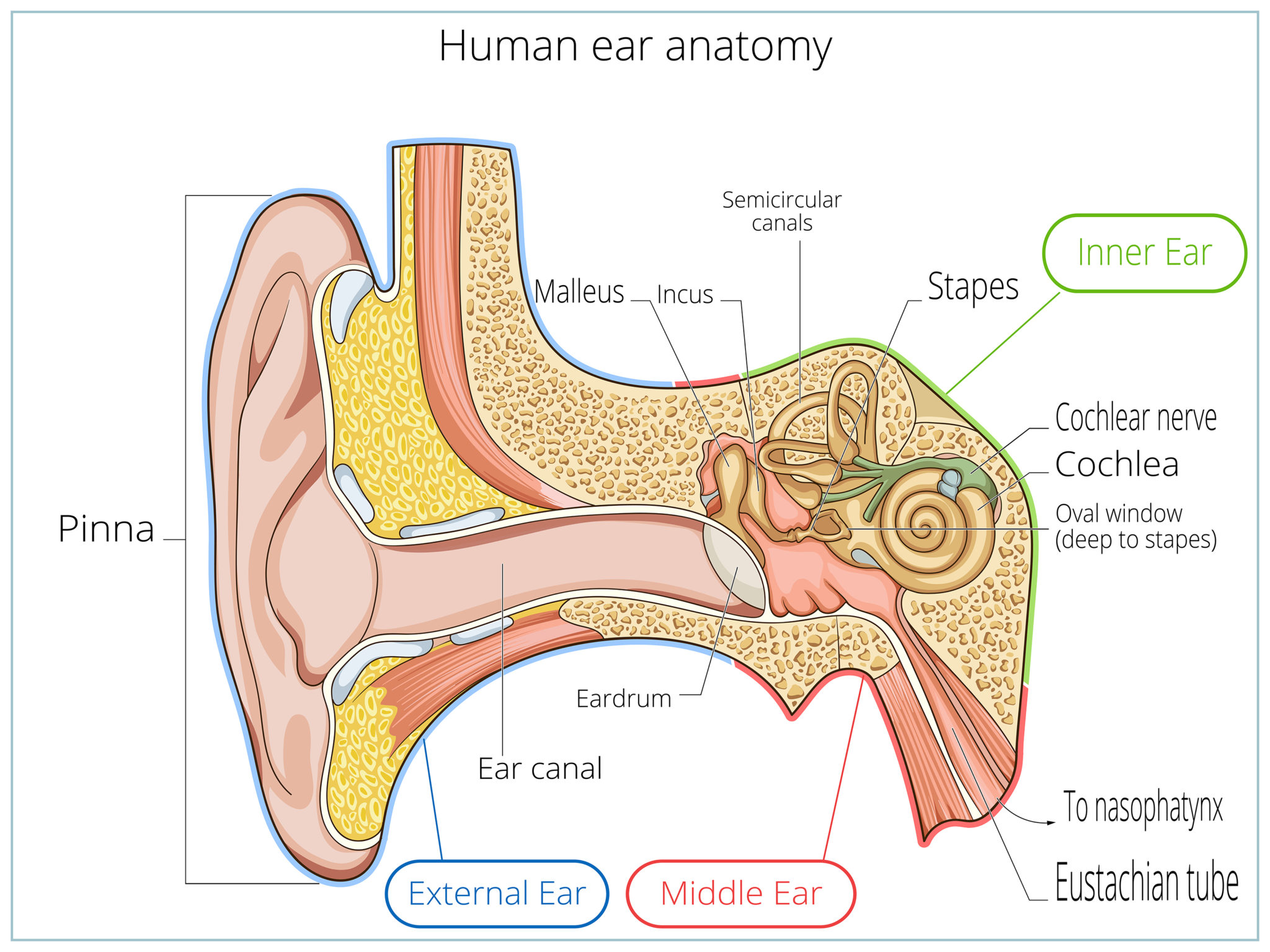
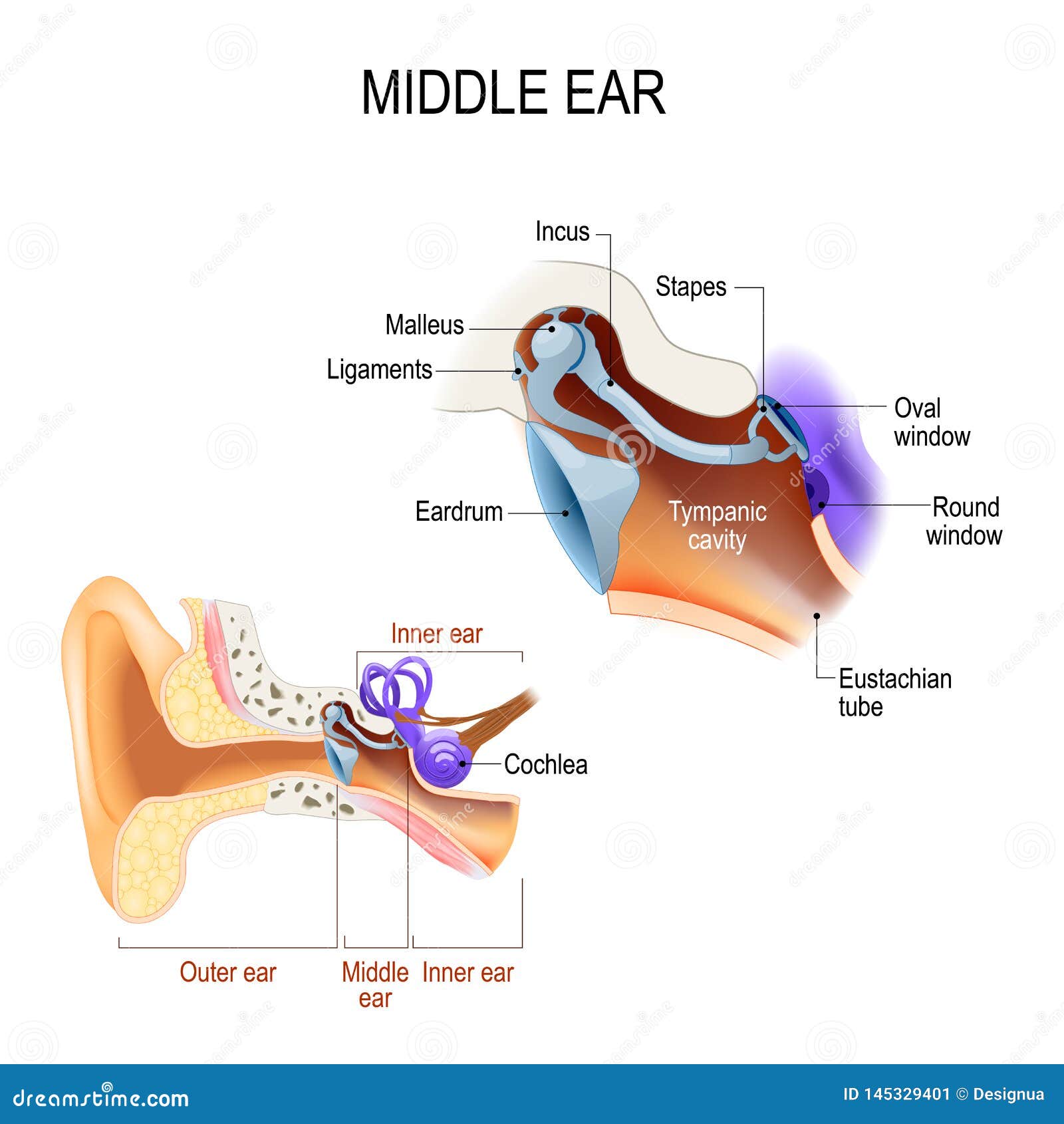
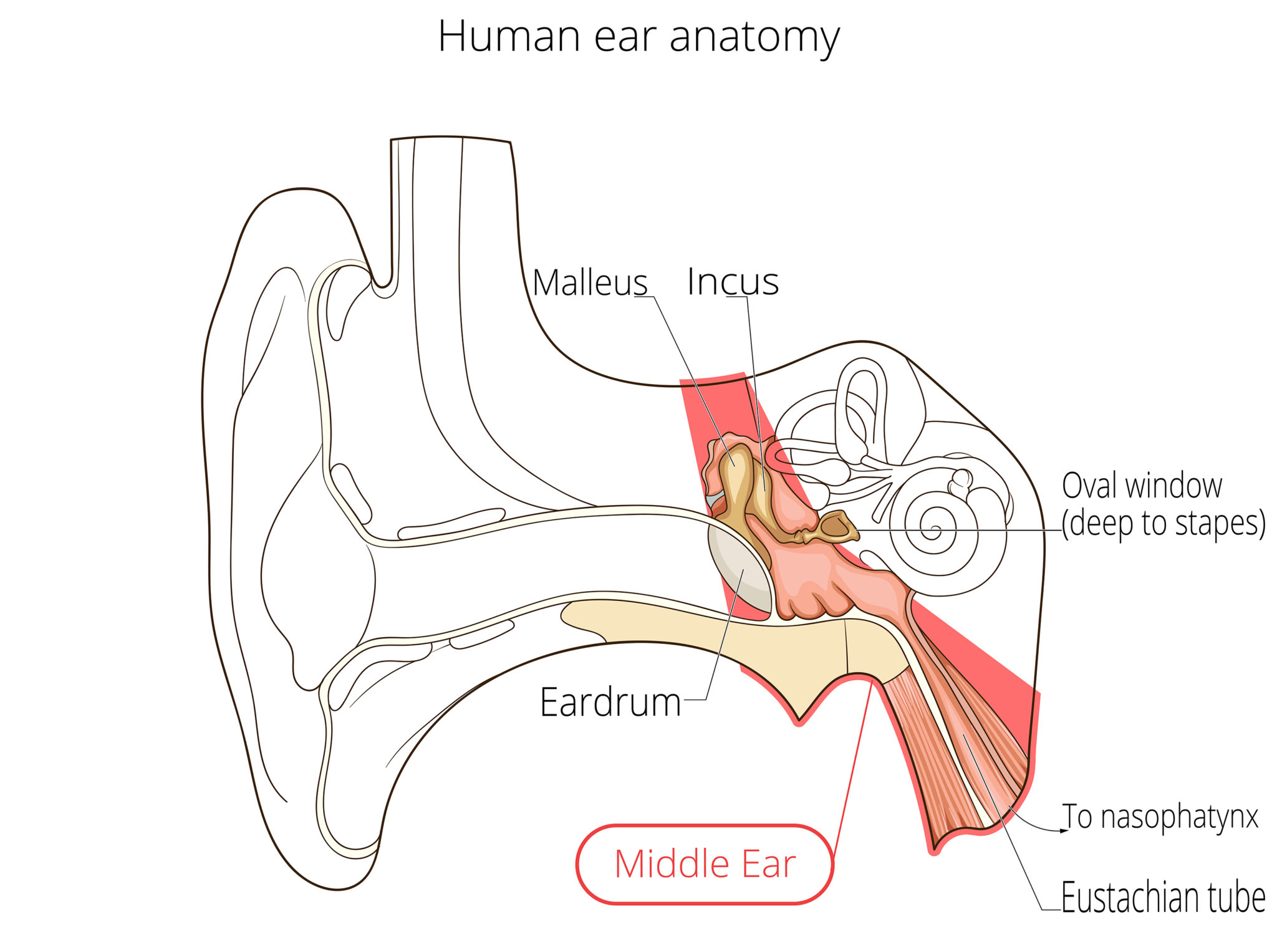
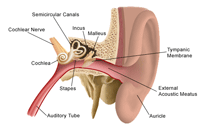
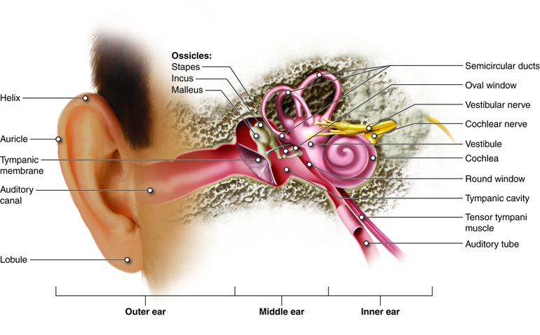
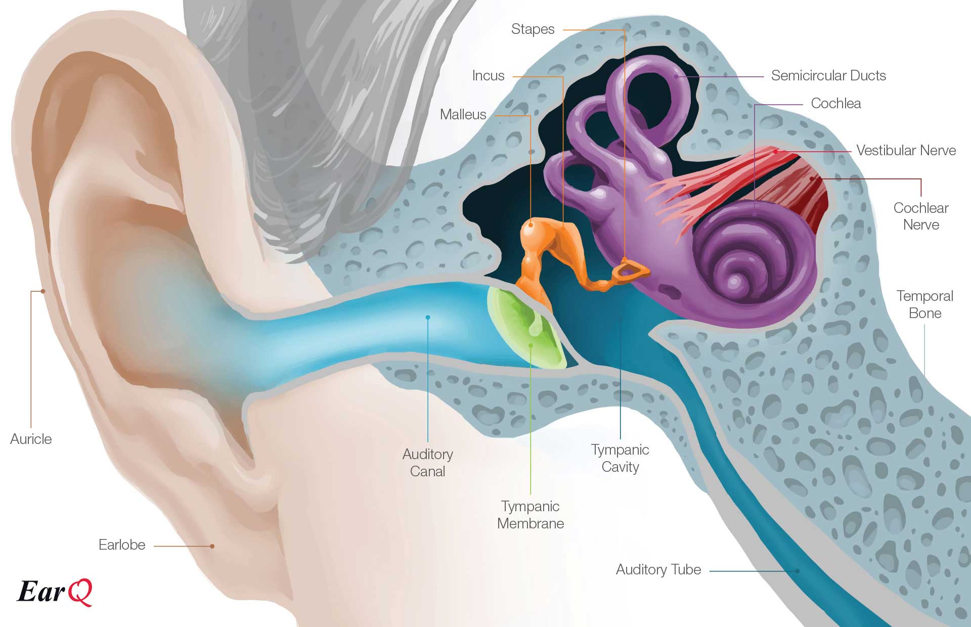
Anatomy of the middle ear. The mammalian middle ear contains three ossicles which transfer the vibrations of the eardrum into waves in the fluid and membranes of the inner ear. The eardrum separates this space from the ear canal. Semicircular ducts filled with fluid.
Three small bones that are connected and transmit the sound waves to the inner ear. A canal that links the middle ear with the back of the nose. Auditory tube drains fluid from the.
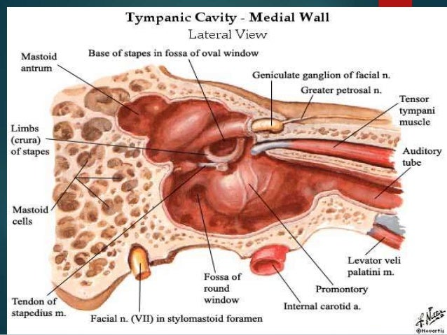
The eardrum acts as a natural boundary between the middle ear and the ear canal. Attached to cochlea and nerves. The hollow space of the middle ear is also known as the tympanic cavity and is surrounded by the tympani bone.
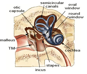
The middle ear consists of three bones. The middle ear is the portion of the ear internal to the eardrum and external to the oval window of the inner ear. The hammer malleus the anvil incus and the stirrup stapes the oval window the round window and the eustrachian tube.
The middle ear is the part of the ear between the eardrum and the oval window. Middle ear tympanic cavity consisting of. Oval window connects the middle ear with the inner ear.
The middle ear transmits sound from the outer ear to the inner ear. Cochlea spiral shaped organ of hearing. The middle ear also known as the tympanic cavity or the tympanum is a pneumatized air filled region of the temporal bone that lies just medial to the tympanic membrane ear drum and lateral to the promontory caused by the turns of the cochlea of the ear.
Transforms sound into signals that get sent to the brain. Also known as the tympanic cavity the middle ear is an air filled membrane lined space located between the ear canal and the eustachian tube cochlea and auditory nerve. The auditory tube joins the tympanic cavity with the nasal cavity allowing pressure to equalize between the middle ear an.
The bones are called. What is the middle ear.
![]() Middle Ear Anatomy Relating Structures And Supply Kenhub
Middle Ear Anatomy Relating Structures And Supply Kenhub
Middle Ear
 Inner Structure Of Human Ear Ear Internal Structure Anatomy
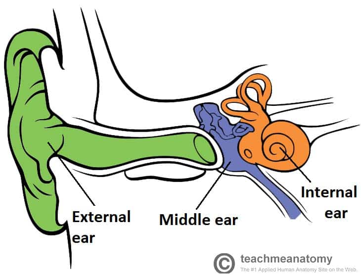
 The Middle Ear Parts Bones Muscles Teachmeanatomy
 Middle Ear Anatomy Notes On Neet Pg
 Bountiful Layton Ear Tube Surgery For Middle Ear Infection
 Middle Ear Anatomy
 Chronic Ear Infections Eastern Virginia Medical School
 Middle Ear Anatomy Springerlink
 Middle Ear Conditions Anatomical Chart
 Middle Ear Three Ossicles Malleus Incus And Stapes
 Middle Ear Anatomy At Grand Valley State University Studyblue
 Raleigh Nc Ear Surgeons And Surgery Patient Education
Anatomy Of Middle Ear Download Scientific Diagram
 Inner Ear Anatomy Function And Health
 Unit One Normal Anatomy
 Anatomy Middle Ear
 Oval Window Wikipedia
 Perilymph Fistula
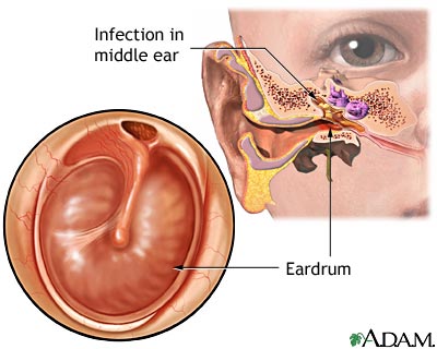
 Otitis Media Middle Ear Infection
 Hearing And Equilibrium Anatomy And Physiology
 The Middle Ear Parts Bones Muscles Teachmeanatomy
 Ear Infection Chronic Medlineplus Medical Encyclopedia
 Anatomy Of The Inner Ear Anatomical Chart
 Anatomy Of The Ear Inner Ear Middle Ear Outer Ear


Posting Komentar
Posting Komentar