The heart is at the center of this system as it pumps blood through vascular channels towards the target tissue. Contains the sinoatrial node.
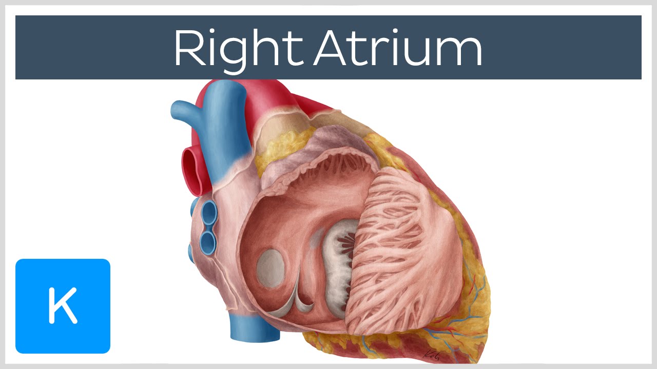 Right Atrium Location Anatomy Function Human Anatomy Kenhub
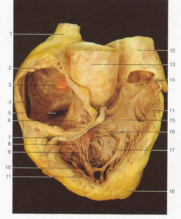
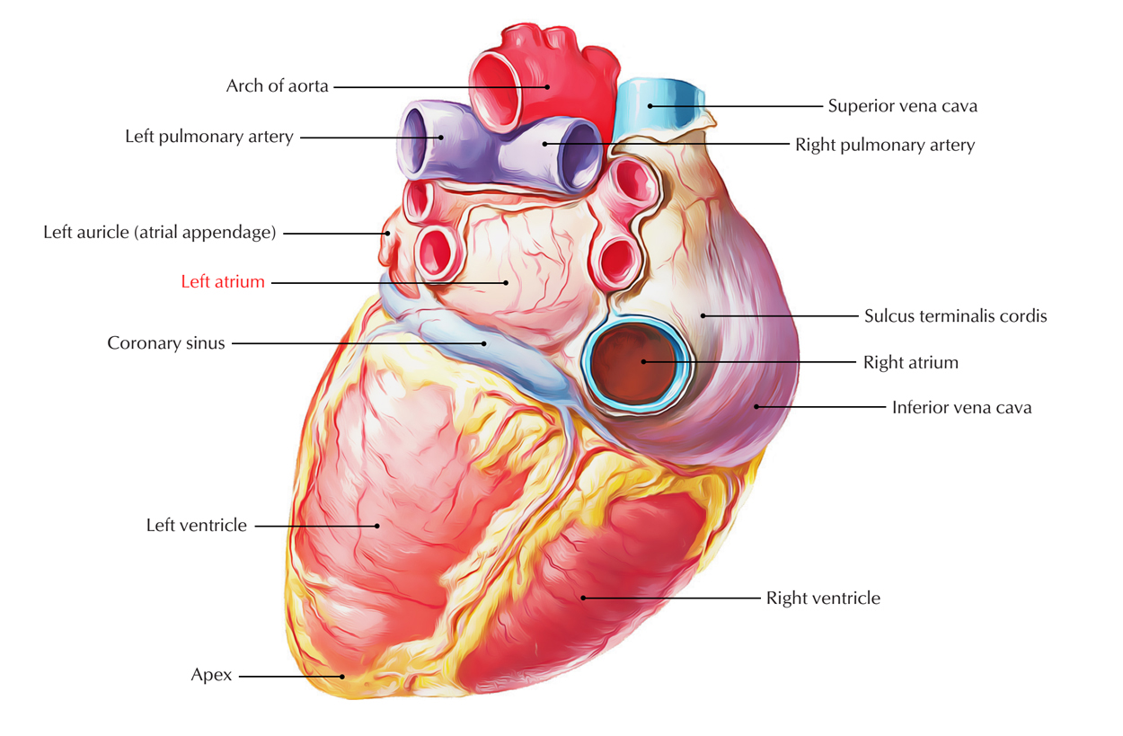
The superior vena cava inferior vena cava and coronary sinus figures 1 and 2.
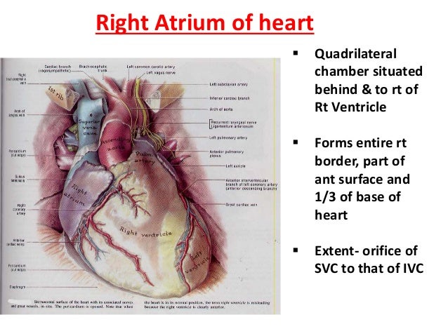
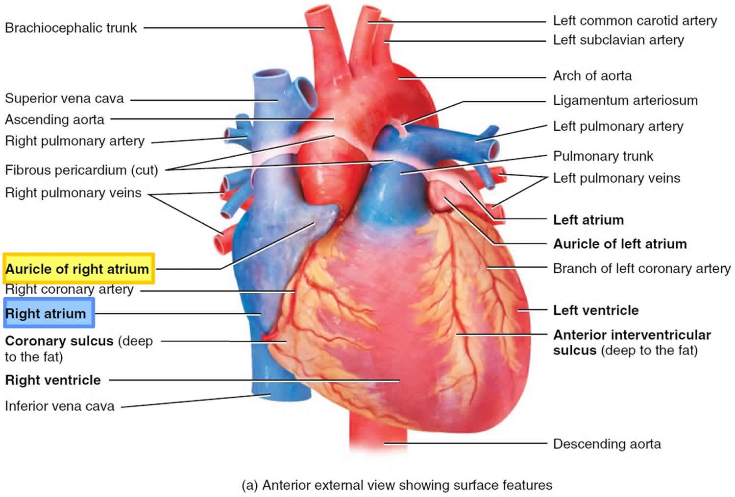
Right atrial anatomy. The right side of the heart then pumps this deoxygenated blood into the pulmonary arteries around the lungs. The anterior right atrial arteries araa originate from the right coronary artery rca which emerges from the anterior ascending aorta. Recall that the heart is a roughly pyramidal organ made up of two muscular pumps that are connected in series namely the left and right hearteach pump contains an upper chamber that functions as a receptacle for incoming blood called the atrium.
The araa primarily supplies blood to the anterior portion of the right atrium and secondarily supplies the inter atrial groove and part of the left atrium. The right atrium forms the entire right border of the human heart. The heart is comprised of two atria and two ventricles.
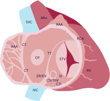
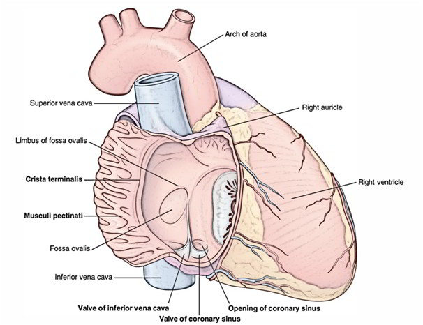
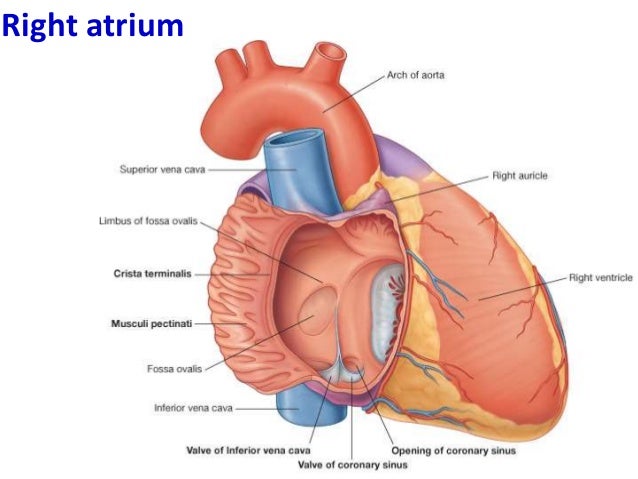
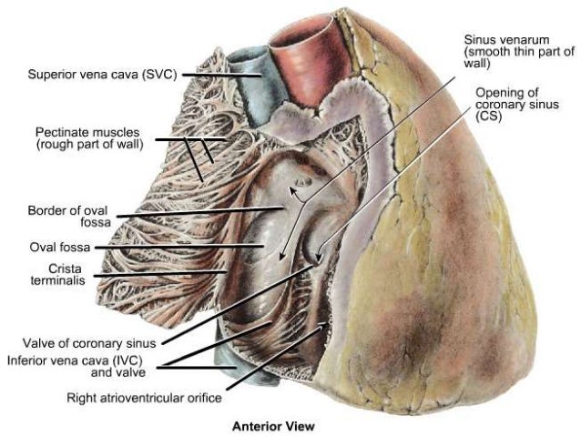
Within the right atrium youve essentially got two spaces. Right atrium gross anatomy. Basic anatomy of the heart.
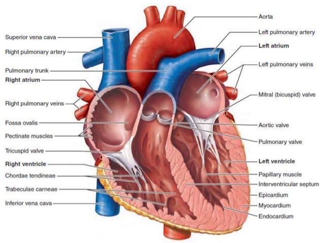
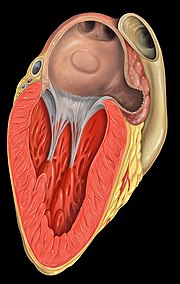
The right atrium is located in the upper portion of right side of heart consisting of the sinus venosus and the right atrial appendage. Deoxygenated blood enters the right atrium through the inferior and superior vena cava. Blood enters the heart through the two atria and exits through the two ventricles.
The right atrium is the location of the sinoatrial node the hearts pacemaker. The right atrium receives oxygen poor blood from three veins. Outflow portion located anteriorly.
Internally this corresponds to the crista terminalis. The right atrium receives deoxygenated blood from the superior vena cava svc. The right atrium is the receiving chamber for oxygen poor blood deoxygenated returning from the systemic circuit.
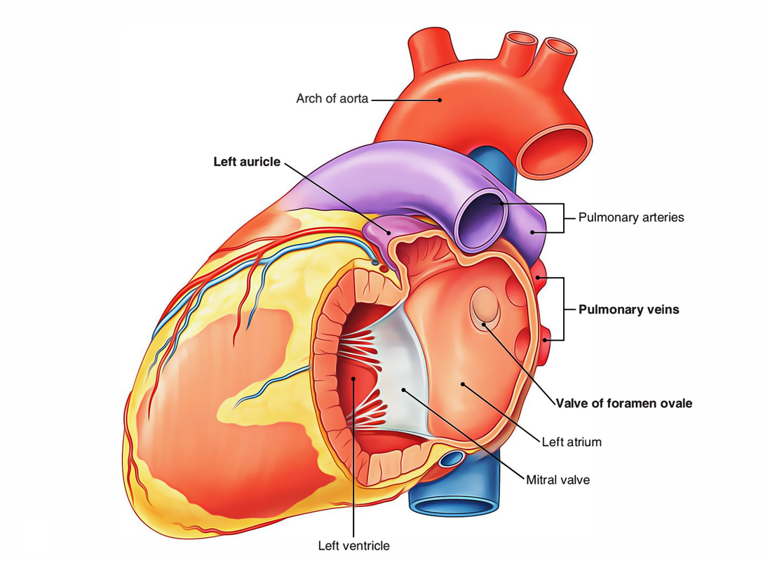
Its internal surface is smooth and it is derived from the pulmonary veins themselves. On contrast enhanced chest ct and. The interior surface of the left atrium can be divided into two parts each with a distinct embryological origin.
Theyre divided by something called the sulcus terminalis on the external surface of the heart. Inflow portion receives blood from the pulmonary veins.
 Left Atrium Earth S Lab
 The Anatomical Distribution Of Ectopic Atrial Tachycardias
 Right Atrium And Right Ventricle
 A Comprehensive Review Of The Anatomical Variations In The
 3 Internal Features Of The Heart
 Left Atrium Earth S Lab
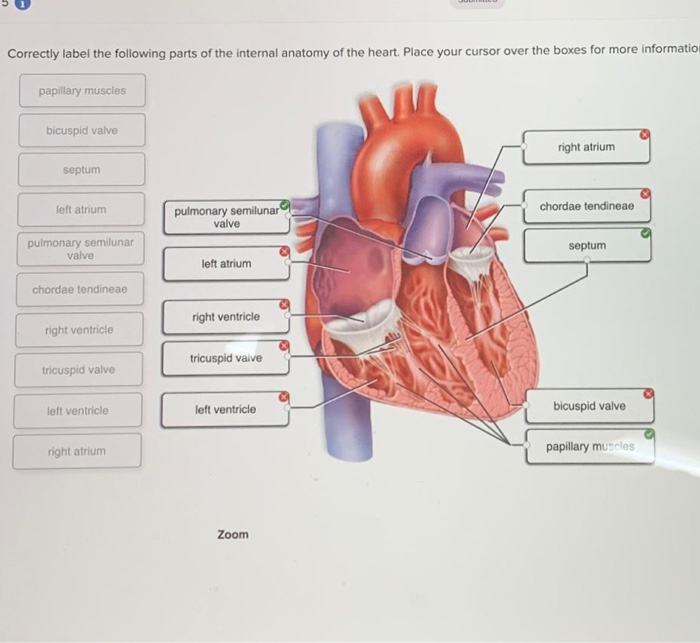 Solved Correctly Label The Following Parts Of The Interna
 Right Atrium And Left Atrium Anatomy Heart Anatomy Drawing
 Difference Between Auricle And Atrium Compare The
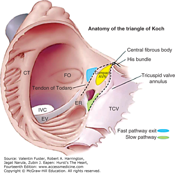 Electrophysiologic Anatomy Hurst S The Heart 14e
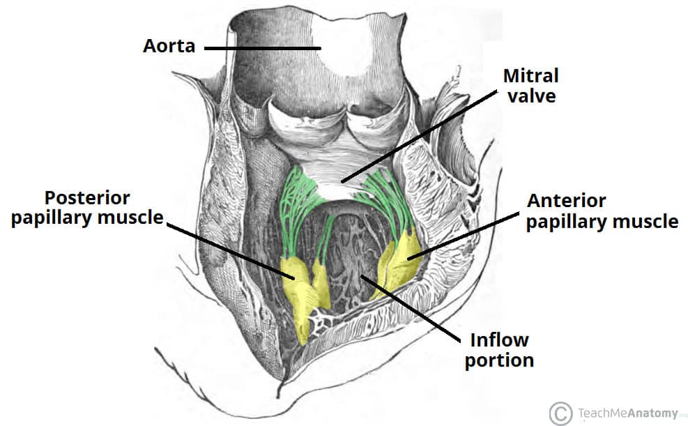 Chambers Of The Heart Atria Ventricles Teachmeanatomy
 Anatomy Of Heart
Cardiac Anatomy
![]() Heart Right And Left Atrium Anatomy And Function Kenhub
Heart Right And Left Atrium Anatomy And Function Kenhub
 Heart Anatomy Chambers Valves And Vessels Anatomy
 3 5 The Right Atrium 123sonography
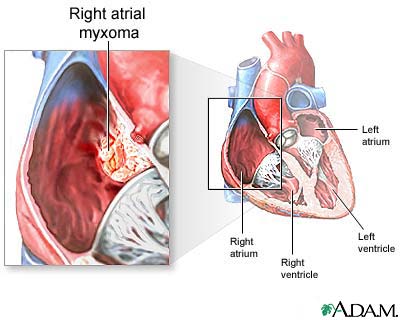
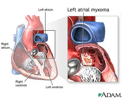 Atrial Myxoma Medlineplus Medical Encyclopedia
 Pediagenosis
![]() Heart Right And Left Atrium Anatomy And Function Kenhub
Heart Right And Left Atrium Anatomy And Function Kenhub
 Right Atrium Location Structure Function Diagram
 Atrium Heart Wikipedia
 Right And Left Atrium Acland S Video Atlas Of Human Anatomy

 Easy Notes On Chambers Of The Heart Learn In Just 3
 3 Internal Features Of The Heart
 Heart Anatomy Opened Right Atrium Right Lateral View
 Anatomy Of The Atria Springerlink
 Right Atrium Anatomy Right Atrium Function Valves
 Interior Of Right Atrium

 Vascular Anatomy And Instrumentation Left Atrium La
 Atrial Myxoma Medlineplus Medical Encyclopedia
 Heart Internal Features Anatomy Qa


Posting Komentar
Posting Komentar