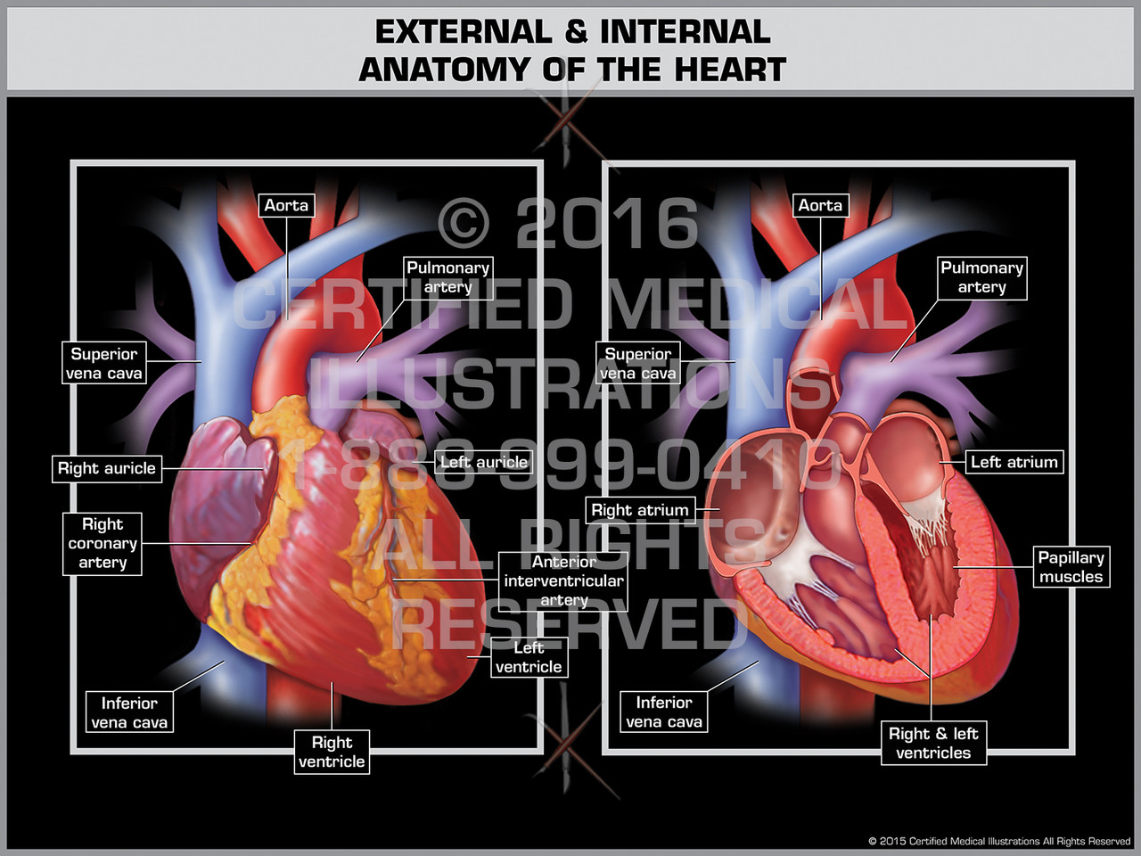
The heart sits within a fluid filled cavity called the pericardial cavity. Auricles are relatively thin walled structures that can fill with blood and empty into the atria or upper chambers of the heart.
 The Heart External And Internal Anatomy
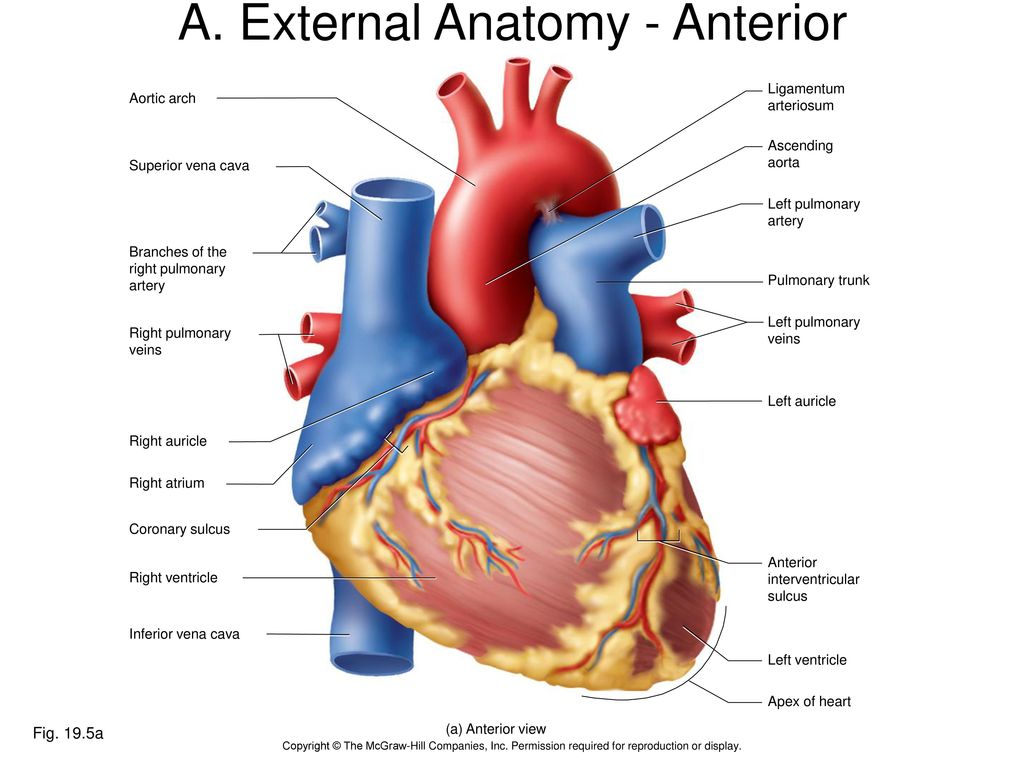
There is a superficial leaf like extension of the atria near the superior surface of the heart one on each side called an auriclea name that means ear likebecause its shape resembles the external ear of a human figure 5.

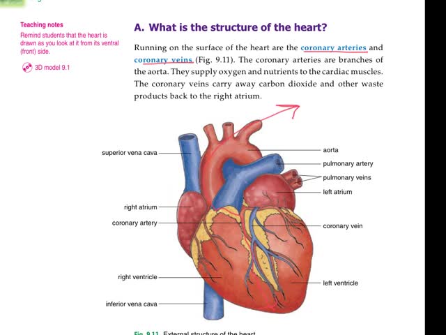
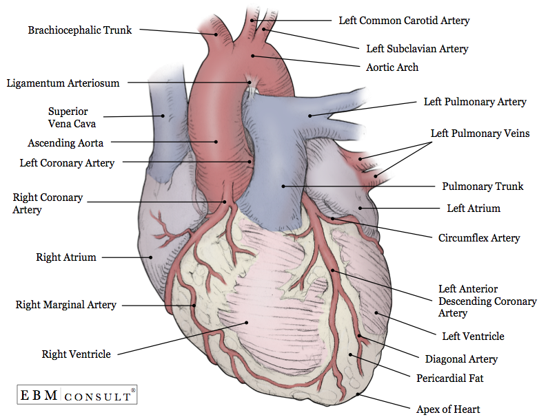
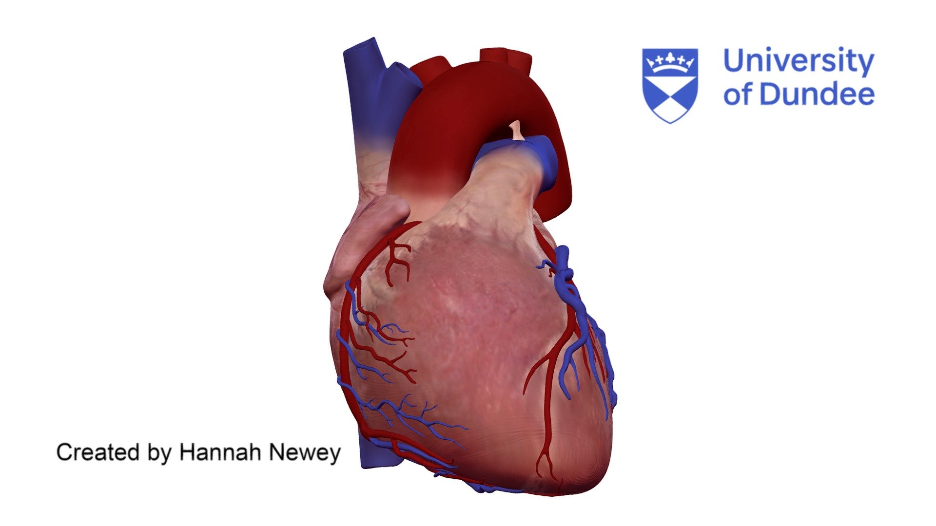
Heart anatomy external. The remainder is supplied by the coronary vasculature which is primarily embedded in the pericardial fat on the surface of the heart and supplies predominantly the epicardium. Heart external anatomy view of the vental surface of the heart. You can identify the front of the heart by locating the interventricular sulcus and the large pulmonary artery.
Anatomy of the heart external and internal structures duration. It uses rhythmic electrical impulses that cause the ventricles to contract and force the blood out of the heart so that the returning blood maybe return in a circular fashion. Apex the distal end of the heart that points downward and to the right coronary artery first.
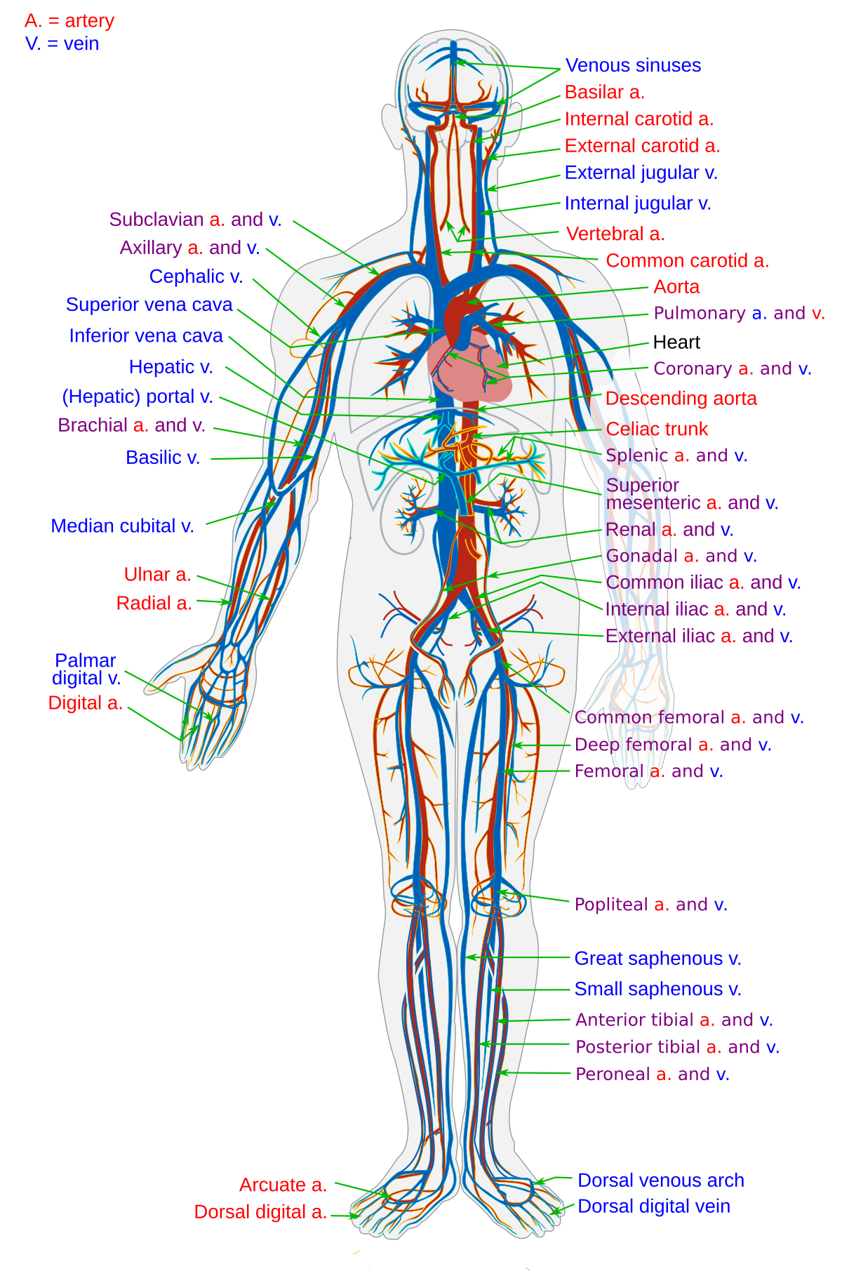
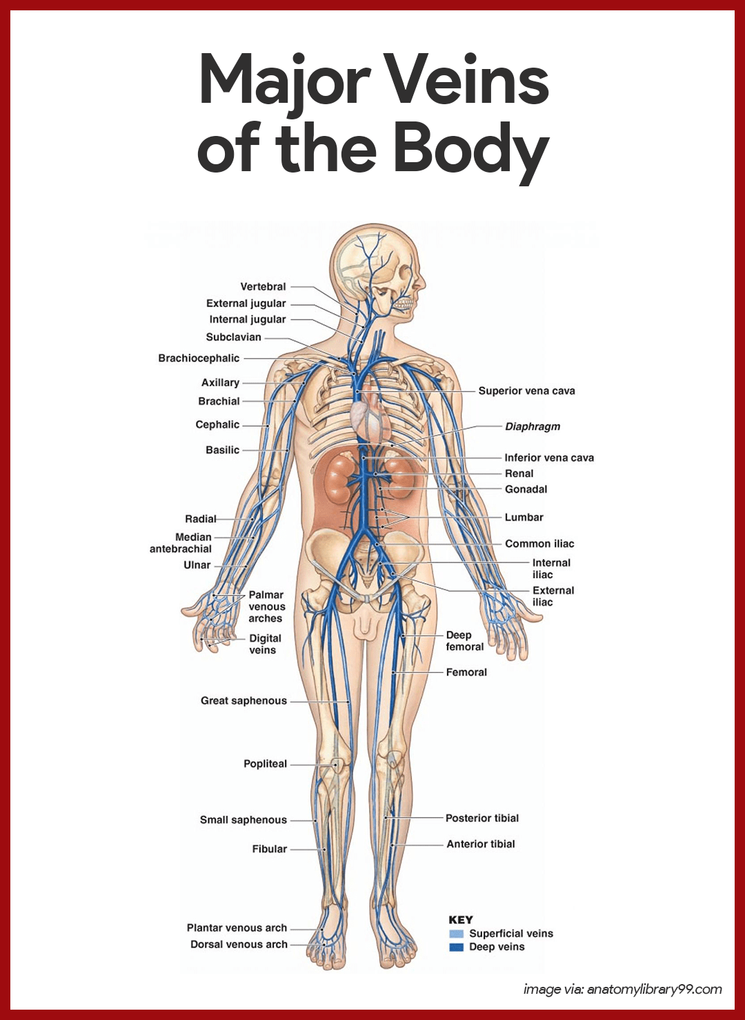
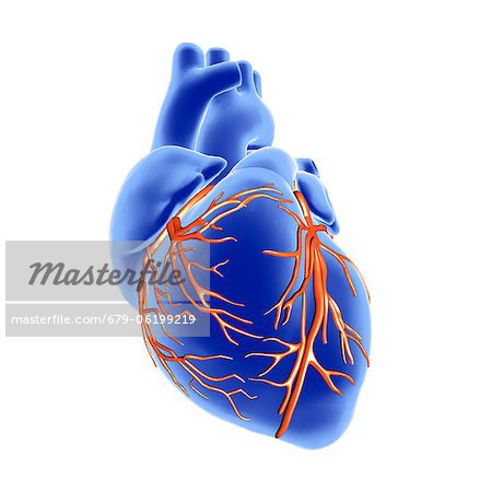
Right coronary artery. Anatomy of the heart pericardium. The heart an image of the heart with blank labels attached the circulatory system upper body image with blank labels attached the circulatory system lower body image with blank labels attached the circulatory system a pdf file of the upper and lower body for printing out to use off line.
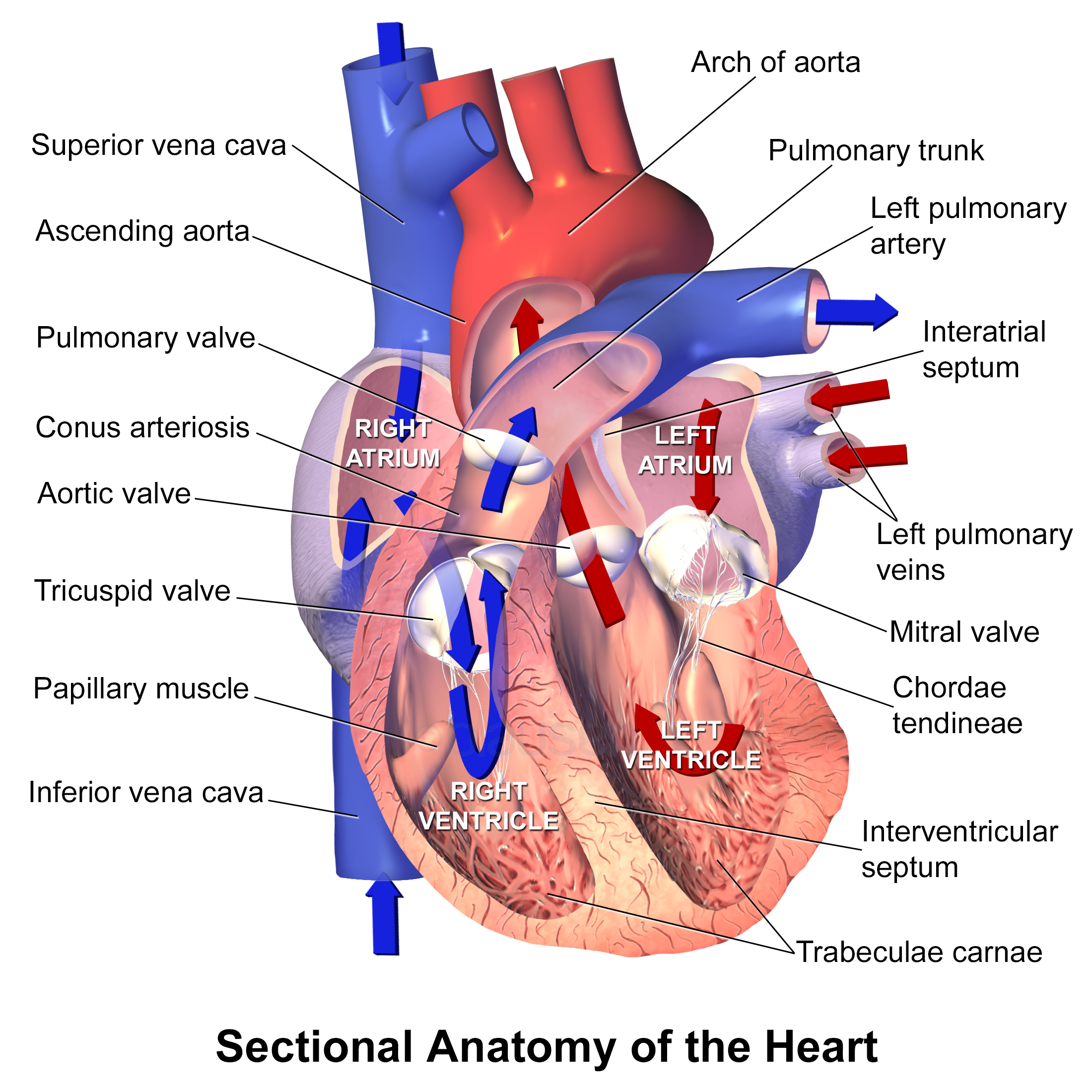
The walls and lining of the pericardial cavity are a special membrane known as the pericardium. In this article the major anatomy of the heart will be discussed such as the borders surfaces the chambers and the great vessels. Apex the distal end of the heart that points downward and to the right coronary artery first arteries to branch off of the aorta.
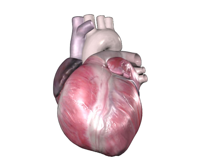
Superior vena cava. The heart is a muscular organ about the size of a fist located just behind and slightly left of the breastbone. Heart anatomy external the endocardium and subendocardial tissue receive oxygen and nutrients by diffusion or microvasculature directly from the chambers of the heart.
A trivia quiz called external heart anatomy. Test your knowledge about external heart anatomy with this online quiz. Because the heart points to the left about 23 of the hearts mass is found on the left side of the body and the other 13 is on the right.
The right one.
 The Human Heart External And Internal Structure Online
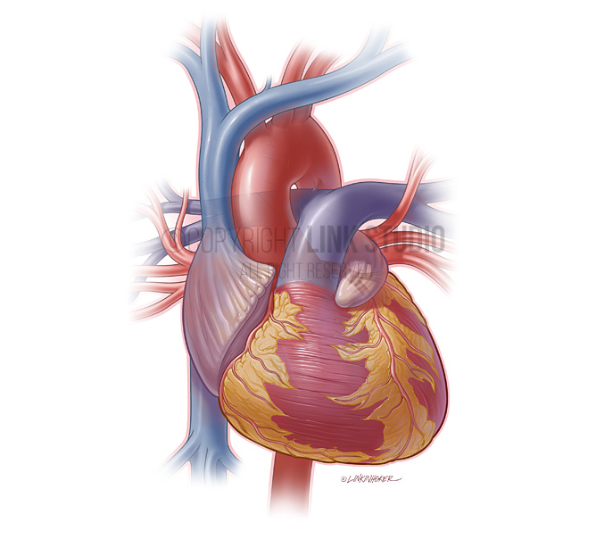 Heart Anatomy Link Studio
Anatomy Of The Heart Medical Illustration Human Anatomy
 The Heart And Cardiac Cycle Lecture Support
 Amazon Com Scientific Human Heart Anatomy Model Enlarged To
 The Heart Internal External Structures Anatomy And Cell
 External Heart Anatomy Posterior View Diagram Quizlet
 Overview Of The Heart Anatomy A Illustration Of The
The Cardiovascular System The Heart
 External Internal Anatomy Of The Heart
 Heart External Features Anatomy Qa
 Blood Vessel Wikipedia

 Anatomy Heart External
 Cardiovascular System Anatomy And Physiology Study Guide
 Hybrid Labeling Of Heart Anatomy With Internal And External
 Cardiac Anatomy External View Of Human Heart Download
 Chapter 19 The Circulatory System The Heart Ppt Download
 Heart And Coronary Arteries 3d Computer Artwork Of The
 2 External Features Of The Heart

 Activity 1 Gross Anatomy Of The Human Heart And Using The
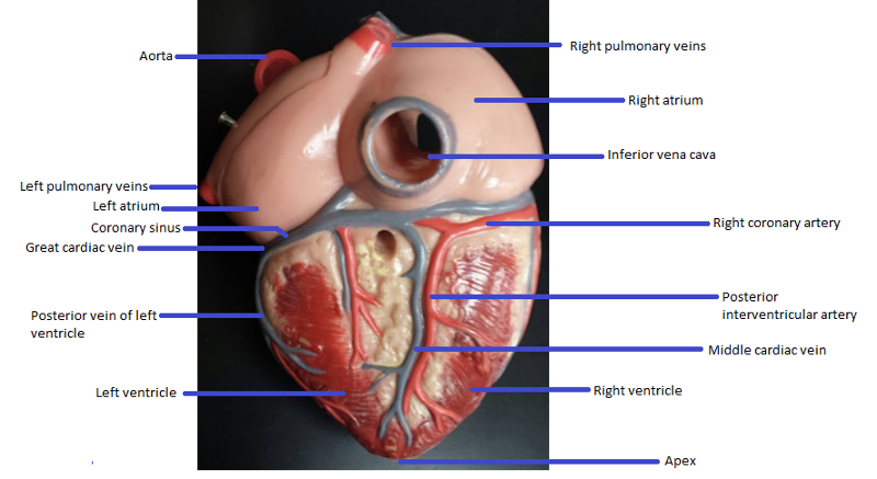
 External Gross Anatomy Of The Heart Posterior View
 1 Anatomy And Histology Of The Cardiac Conduction System In
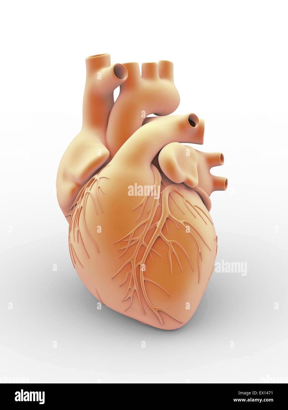 Heart And Coronary Arteries Artwork Of The External Anatomy


Posting Komentar
Posting Komentar