The muscle originates from the body of the pubis and attaches to the pectineal line and proximal part of the linea aspera of femur. Pelvic xrays are a key component of trauma fractures and dislocations seen every day in the ed but when is the last time you went back over the anatomy and radiographic tips and tricks of the pelvic radiograph.
 The Pelvis And Hip
Ilium ischium and pubis connected by the triradiate cartilage.

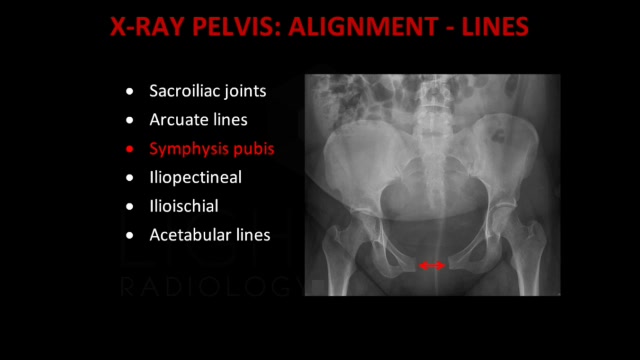
Pelvis xray anatomy. See an approach to the pelvic radiograph. Explore over 5400 anatomic structures and more than 375 000 translated medical labels. The symphysis pubis joint space should be 5 mm.
E anatomy is an award winning interactive atlas of human anatomy. The pelvis series examines the main pelvic ring obturator foramina sacroiliac joints symphysis pubis acetabulum sacral foramina and the proximal femur. Until puberty each hip bone consists of three separate bones yet to be fused.
48 adductor longus muscle this muscle is the most anterior of the adductor group of muscles in the thigh. Mands thorough break down of this commonly used ed diagnostic the pelvic xr. It is enervated by the obturator nerve.
As with other anatomical bone rings if a fracture is seen in one place a careful check should be made for a second fracture or for disruption of the pubic symphysis or sacroiliac joints. Its primary function is the transmission of forces from the axial skeleton to the lower limbs as well as supporting the pelvic viscera. If either joint space is widened think main pelvic ring fracture.
Pelvis judet view the oblique pelvis otherwise known as the judet view is an additional projection to the pelvic series when there is suspicion of an acetabular fracture. The ap pelvis has a diagnostic yield of 94 in severely injured patients 2 3. The judet view is comprised of two projections first the iliac oblique for assessment of the posterior column and anterior wall of the acetabulum.
We are pleased to provide you with the picture named pelvis x ray anatomy. The bony pelvis comprises the two hemi pelvis bones which are bound anteriorly at the pubic symphysis and posteriorly at the sacroiliac joints. Pelvis x ray anatomy in this image you will find the sacroiliac joint acetabular obturator foramina greater trochanter pubic symphysis femoral heads lesser trochanters in it.
Ct mri radiographs anatomic diagrams and nuclear images. Entirety of the bony pelvis is imaged from superior of the iliac crest to the proximal shaft of the femur obturator foramina appear equal iliac wings have an equal concavity greater trochanters of the proximal femur are in profile. It is the most complete reference of human anatomy available on web ipad iphone and android devices.
The sacroiliac joints should be symmetrical joint space range 2 4 mm.
 Amazon Com Ahawoso Seasonal Garden Flag 12x18 Inches

 Radiography Of The Skeleton All Anatomical Structures
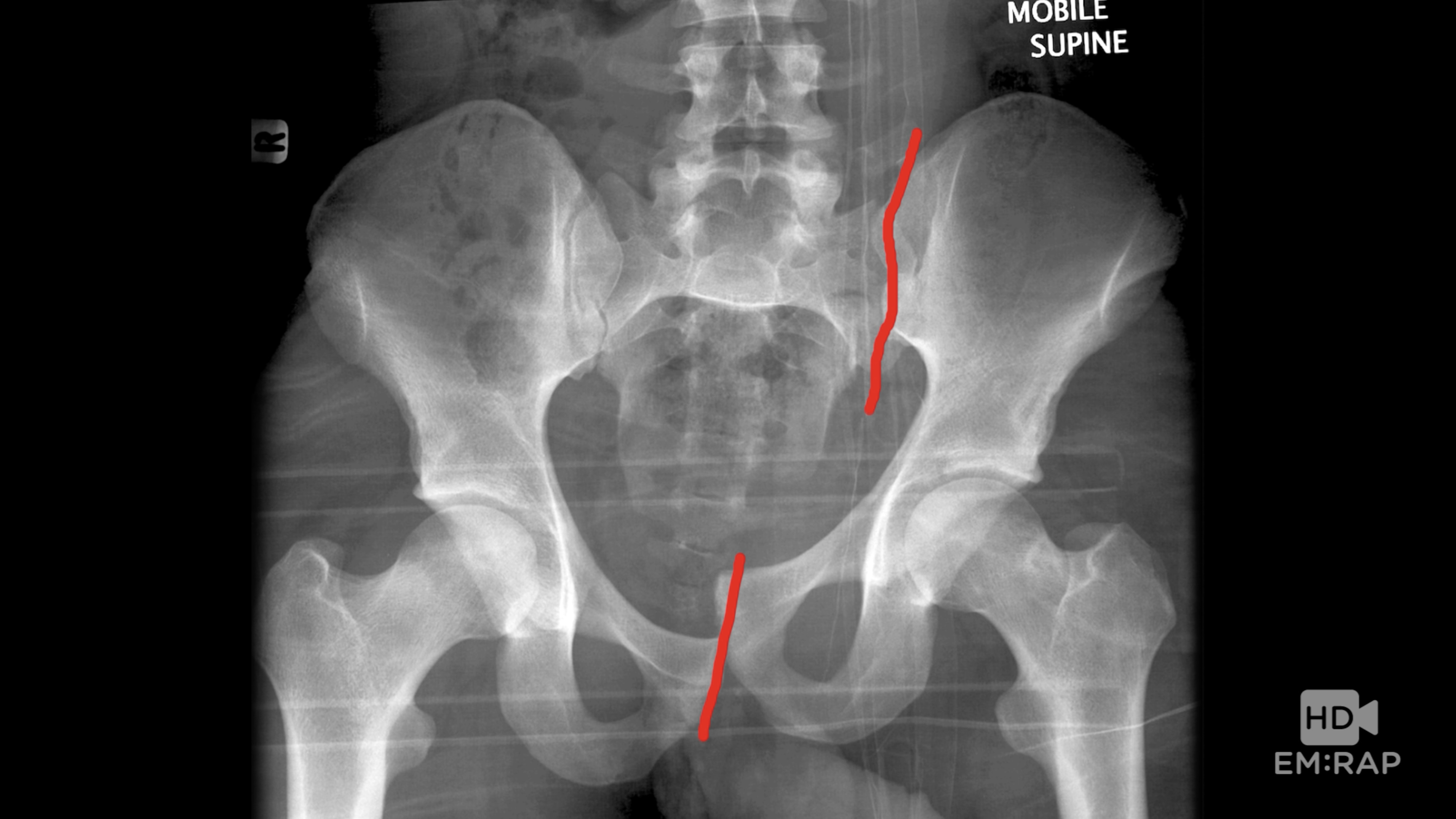 Hd Pelvic Fractures Em Rap
 Skeletal Trauma
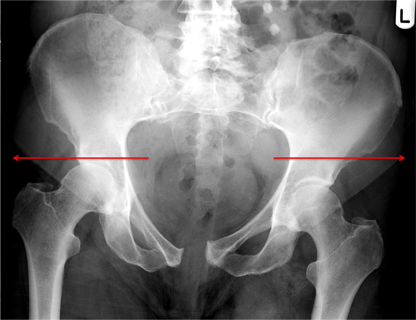
 Anatomical Lines Of The Pelvis On An Anterioposterior
 Pelvic Ring Fractures Trauma Orthobullets
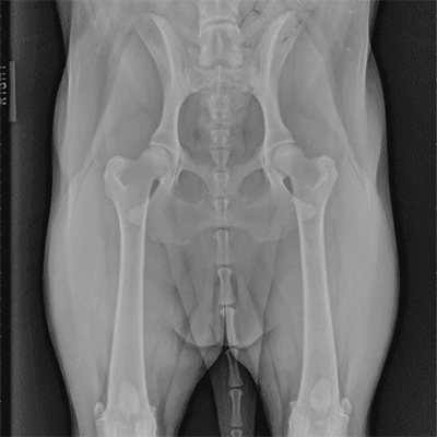
Small Animal Radiography Stifle Joint And Crus Today S
 Film Critique Of The Lower Extremity Part 1

 Pelvis X Ray Ap View Showing Left Sided Dysplastic Hip

 Imaging For Nonarthritic Hip Pathology Mdedge Surgery

 Royalty Free Female Pelvis Stock Images Photos Vectors
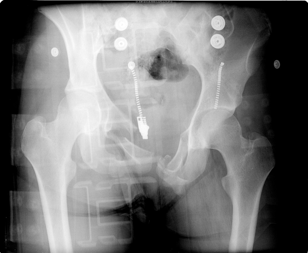
 Pelvis Bone Anatomy X Ray Scan
 The Pelvis And Hip
 Imaging Anatomy
 How To Read Pelvic X Rays International Emergency Medicine
 Pelvis
 Ilium Bone Hip Bone Image Photo Free Trial Bigstock

 Emergency Radiography The Bmj
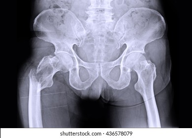
![]() X Ray Image Of Human Normal Spine Rips Pelvis Both Hip
X Ray Image Of Human Normal Spine Rips Pelvis Both Hip


Posting Komentar
Posting Komentar