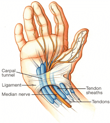
The carpal tunnel is narrowed as a result usually from swelling. In the human body the carpal tunnel or carpal canal is the passageway on the palmar side of the wrist that connects the forearm to the hand.
Basically the carpal tunnel is a passageway for the tendons and nerves of the forearm to travel into the wrist and hand.
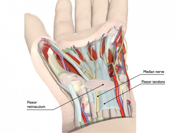
Anatomy of carpal tunnel. These tissues are called the synovium. It serves as the entrance to the palm for several tendons and the median nerve. Gross anatomy boundaries superficial border roof.
Risk factors include obesity repetitive wrist work pregnancy genetics and rheumatoid arthritis. In this article we will look at the borders and contents of the carpal tunnel and its clinical significance. The carpal tunnel is a fibro osseous canal that acts as a passageway from the forearm to the anterior hand.
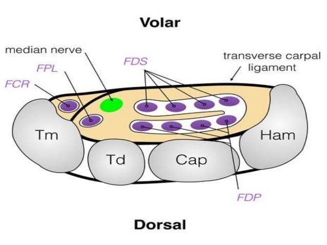
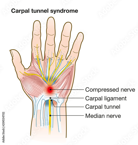
The flexor pollicis longus the four flexor digitorum superficialis the four flexor digitorum profundus as well as the median nerve. Carpal tunnel syndrome occurs when the tunnel becomes narrowed or when tissues surrounding the flexor tendons swell putting pressure on the median nerve. There are described cases of variable median artery occurrence.
Tenosynovitis inflammation of synovial sheaths of long flexor tendons. It is caused by compression of the median nerve in the carpal tunnel. Flexor retinaculum deep border floor.
The canal is narrow and when any of the nine long flexor tendons passing through it swell or d. The flexor pollicis longus has its own synovial sheath whereas the flexor digitorum superficialis and profundus have a common synovial sheath. Normally the synovium lubricates the tendons making it easier to move your fingers.
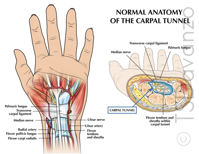
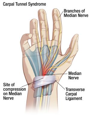
Causes for compression of median nerve in the carpal tunnel can be. Its caused by pressure on your median nerve which runs the length of the arm goes through a passage in the wrist called the carpal tunnel and ends in the hand. The carpal tunnel is a narrow passageway found on the anterior portion of the wrist.
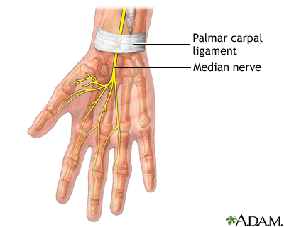
Anatomy of the carpal tunnel showing the median nerve passing through the tight space it shares with the finger tendons most cases of cts are of unknown cause. Carpal tunnel anatomy a passageway from the wrist to the hand the carpal tunnel is made of tendons ligaments and bones. The median nerve passes through the tunnel and provides sensation to your thumb index finger middle finger and the thumb side of the ring finger.
The median controls the movement and feeling of your thumb and also the movement of all your fingers except your pinky. The carpal tunnel contains nine tendons and a nerve. Osteoarthritis involving carpal bones.
The walls of the tunnel are formed by the bones and ligaments of the wrist while the tendons and nerves travel through the tunnel and are protected from external forces by the bones and ligaments. Fluid retention in pregnancy. Dislocation of lunate bone.
The tunnel is bounded by the bones of the wrist and flexor retinaculum from connective tissue. Normally several tendons from the flexor group of forearm muscles and the median nerve pass through it. Diabetes mellitus is weakly associated with cts.
 Left Carpal Tunnel And Guyon S Canal Release
 Anatomical Model Wrist Hand Carpal Tunnel Syndrome Science


 Carpal Tunnel Treatment By Omaha Hand Doctors Md West One
 Weekend Wellness Carpal Tunnel Affects Hands Symptoms
 Hand Anatomy Google Search Hand Anatomy Median Nerve
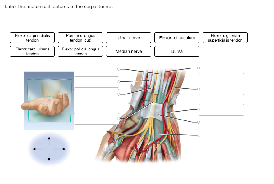 Solved Label The Anatomical Features Of The Carpal Tunnel
 Carpal Tunnel Syndrome Lehigh Valley Health Network
 Open Techniques For Carpal Tunnel Release Musculoskeletal Key
 Open Techniques For Carpal Tunnel Release Musculoskeletal Key
 Patient Basics Carpal Tunnel Syndrome 2 Minute Medicine
![]() Carpal Tunnel Syndrome Durrant Orthopaedics
Carpal Tunnel Syndrome Durrant Orthopaedics
 Carpal Tunnel Syndrome Upswing Health
 Hand And Wrist Anatomy Baxter Regional Medical Center
 Carpal Tunnel Syndrome Anatomy Medical Illustration With
 Carpal Tunnel Repair Series Normal Anatomy Medlineplus
 Trialsight Exhibits Slaybaugh Studios
 Carpal Tunnel Syndrome Picture Image On Medicinenet Com

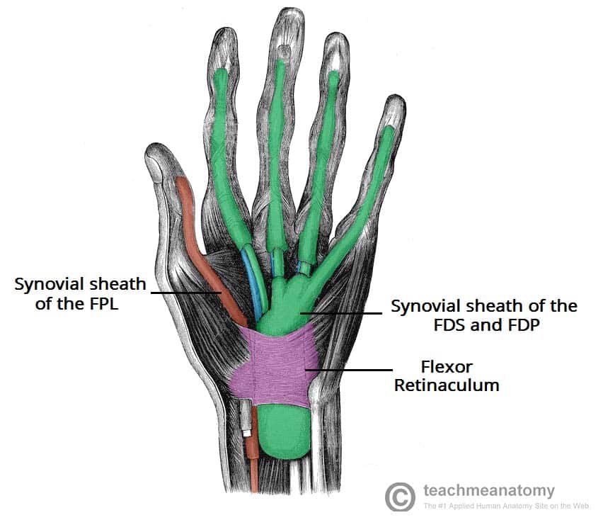 The Carpal Tunnel Borders Contents Teachmeanatomy
 Carpal Tunnel Cedar Park Tx Austin Tx Hand Surgery
 Mayo Clinic Q And A Recovery After Surgery For Carpal
Carpal Tunnel Syndrome Anatomy And Radiology Imaging Findings
 Carpal Tunnel Syndrome
 Carpal Tunnel
 Flexor Retinaculum Of The Hand Wikipedia


Posting Komentar
Posting Komentar