It also includes the joints of the hip stifle hock fetlock pastern and coffin. That way if you need to talk to a vet or do a correct drawing youll have a solid foundation.
 Musings At Minkiewicz Studios Llc Equine Anatomy And
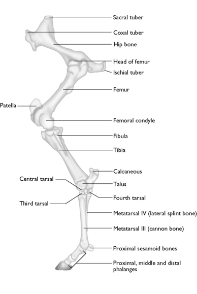
After the pelvis come the femur thigh patella stifle joint tibia fibula tarsal hock bone and joint large metatarsal cannon and small metatarsal splint bones.

Horse hind leg anatomy. The tarsus is the corresponding joint on the hind leg commonly called the hock. Directional terms skeletal and muscle introduction. The horses knee is one of the most complex regions in the limb because there are several small bones and ligaments all combining to form the three main joints.
It looks as if the horse is taking very high steps with the back legs. The foot of the horse. These horse anatomy diagrams are a great overview and introduction to the vast study of equine anatomy.
They are joined to the spine through the sacroileac joints and allow transfer of propulsion to the hind legs. Stringhalt is the over flexing of one or both back legs. This ideal design maximizes the hindlegs power to move the body forward.
Unlike the front leg the hindleg is directly attached to the spine via the pelvis. Each hind limb of the horse runs from the pelvis to the navicular bone. The ischium forms the point of the buttock.
It also includes the joints of the hip stifle hock fetlock pastern and coffin 19 the stifle is the largest single joint in the body. The flexing can be more subtle however appearing occasionally and can be more obvious when the horse is asked to step back or turn sharply. The hindleg together with the frontleg forms the appendicular skeleton of the horse.
The tarsus of the horse hindlimb equivalent to the human ankle and heel the large joint on the hind leg. The horse leg anatomy in the rear includes the bones of the pelvis the ilium ischium and pubic bones femur tibia fibula metatarsus and the phalanxes. The horse leg anatomy in the rear includes the bones of the pelvis the ilium ischium and pubic bones femur tibia fibula metatarsus and the phalanxes.
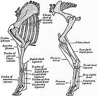
Horse hind leg anatomy sectional view in this image you will find the proximal suspensory ligament deep digital flexor tendon suspensory ligament superficial distal sesamoidean ligament deep digital flexor tendon extensor branch of suspensory ligament in it. The horses hind limbs the top part of the hind limbs consists of three fused bones called the ileum ischium and pubis. Equine anatomy refers to the gross and microscopic anatomy of horses and other equids.
The radiocarpal intercarpal and carpometacarpal joints. The horse will snap the hoof upwards and then stomp down. The hoof wall is the tough outside covering of the hoof that comes into contact with the ground and is.
These diagrams should explain and show you some of the basics. Horse rear leg anatomy horse rear legs.
Equine Hindlimb Regional Joint Bone Anatomy Chart Horse
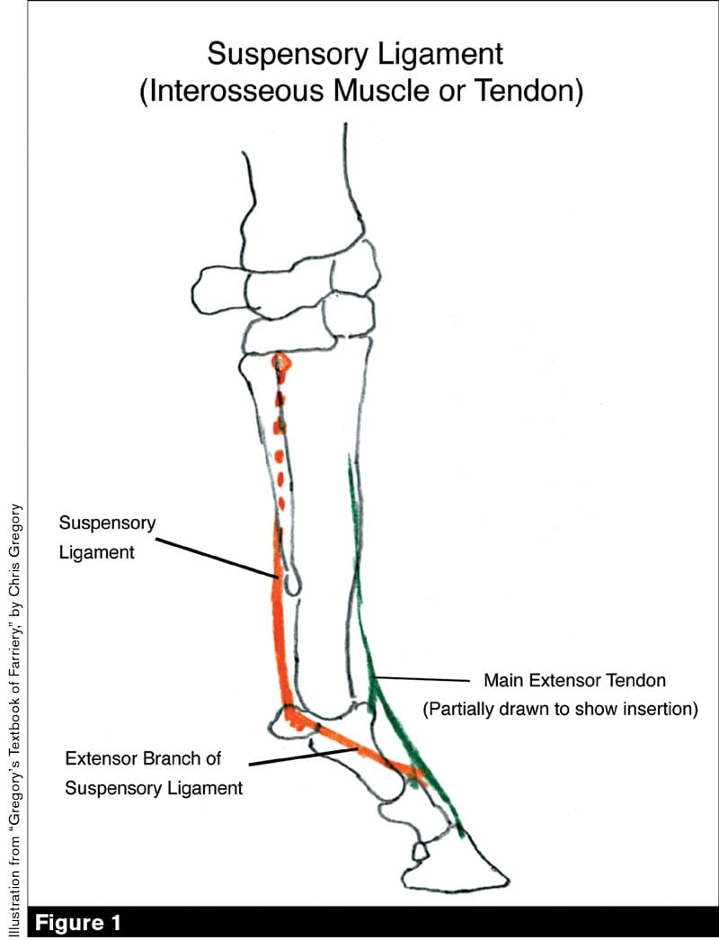 Increased Knowledge Of The Equine Anatomy Can Help Farriers
 Anatomy Of The Horse
 Forever Horses Anatomy Of The Equine Hindleg Horse
 The Horse Veterian Key
 Horse Health At Southern Pines Equine Associates Southern
 Anatomy And Physiology On Equine Art Reference Deviantart
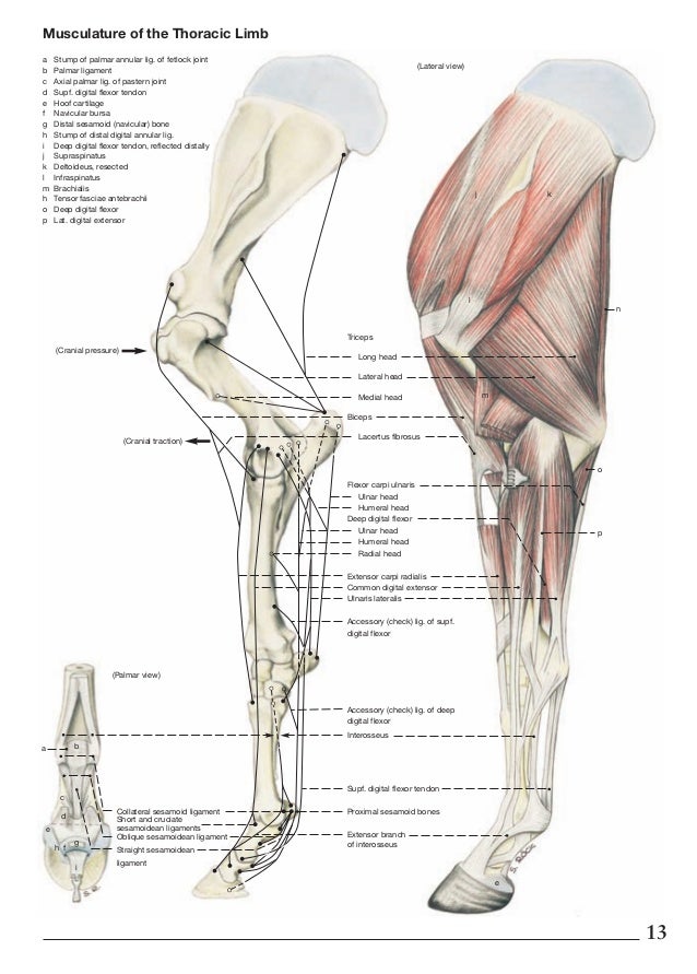
 Comparative Anatomy And Muscle Architecture Of Selected Hind
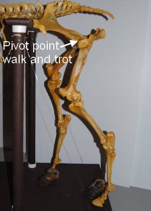 Of Pivots Lollipops And Springs Movement Of The Horse S
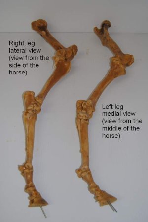 Bones Of The Hind Leg Part One
 The Effect Of Gait And Digital Flexor Muscle Activation On
 Horse Bones Everything You Need To Know To Get Started
 Horse Leg Anatomy Front And Rear Leg Anatomy
 Joint Work And Power For Both The Forelimb And Hindlimb

 Why Do Some Animals Have Forward Facing Front Knees Like
 Suspensory Injuries In Horses Expert How To For English Riders
 Limbs Of The Horse Wikipedia
 Stringhalt The Marionette Horse Irongate Equine Clinic
 Horse Hind Leg Bones Horse Equus Anatomy Isolated On
 Deep Digital Flexor Tendon Injuries Does It Mean The End Of
 Forever Horses Anatomy Of The Equine Hindleg
 Novobrace Tendonitis Desmitis And Soft Tissue Injury
 Leg Anatomy Britannica
 Causes Of Equine Lameness Equimed Horse Health Matters
 Understanding Your Horse S Hock Health Dressage Today
 Limb Or Limbs Upper Hind
 Horse Leg Anatomy Learn Everything You Did Not Know Medrego
 The Not So Fab Four Diseases Resulting In Hind Limb Gait
 Novobrace Tendonitis Desmitis And Soft Tissue Injury
 Front Leg Locking Mechanisms Horse Anatomy Horses Horse
 Habitat For Horses Kids Corral


Posting Komentar
Posting Komentar