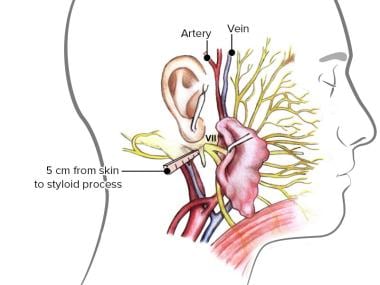
 Facial Nerve Anatomy Overview Embryology Of The Facial
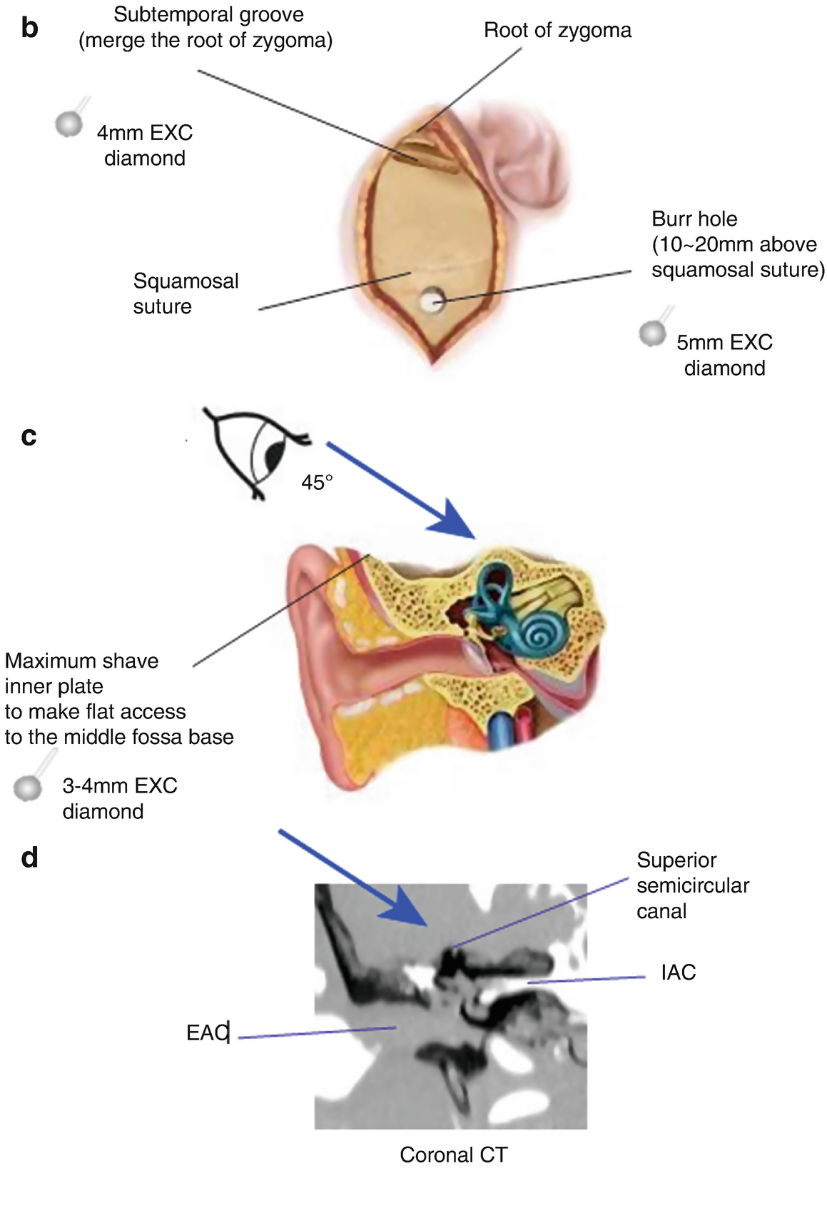 Dissection Of Extended Middle Fossa And Anterior
The Inner Ear Imaging Anatomy With 3t Mri New Sequences A
 A C Axial Temporal Hrct Images Of Normal Inner Ear Anatomy
 Chapter 61 Vestibular Schwannoma Acoustic Neuroma
 Human Brain Brain Iac Fun Learning Bundle Save 20
The Inner Ear Imaging Anatomy With 3t Mri New Sequences A
Imaging Findings Of Cochlear Nerve Deficiency American
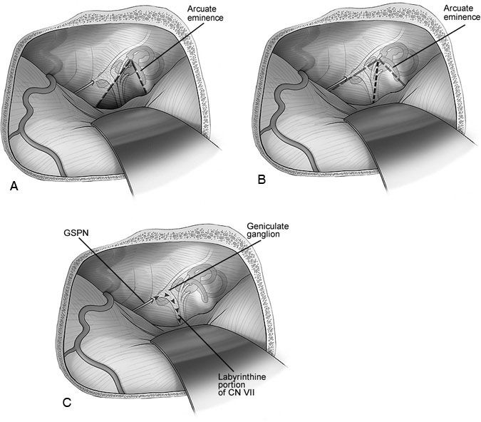 The Middle Fossa Approach Barrow
 Three Dimensional Imaging Of The Human Internal Acoustic
 Normal Mri Internal Auditory Canal Radiology Case
 Figure Measurements Of Internal Auditory Canal Iac A
Surgical Exposure Of The Internal Auditory Canal Through The
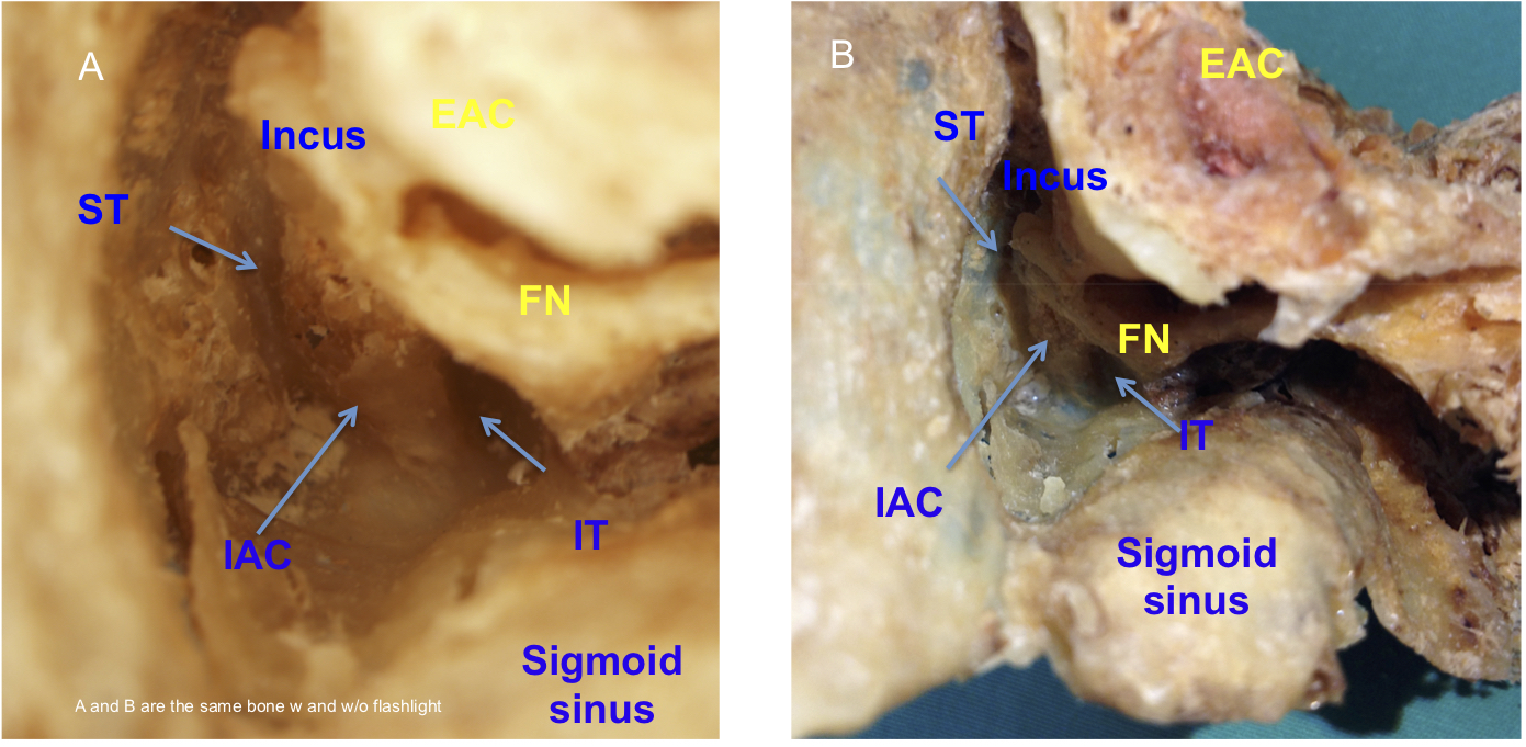 Temporal Bone Anatomy Cadaveric Dissection Iowa Head And
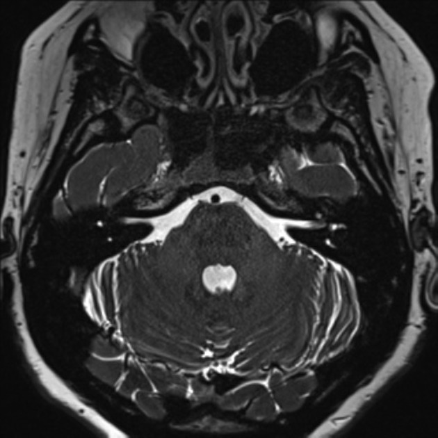 Normal Mri Internal Auditory Canal Radiology Case
 Figure 1 Ct Imaging Of The Anatomy Of The Eustachian Tube
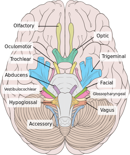 Cranial Nerve Anatomy Cranial Nerves Iowa Head And Neck
 Human Brain Brain Iac Fun Learning By Judy Karim Tpt
 Roentgen Ray Reader Contents Of The Internal Auditory Canal
 A Study Of Middle Cranial Fossa Anatomy And Anatomic
 Cerebellopontine Angle Tumors Ento Key
Cn Vii Facial Nerve Learnneurosurgery Com
 Classification Of The Position And Relationship Of The Iac
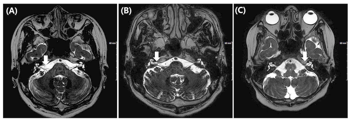 Anatomical Location Of Aica Loop In Cpa As A Prognostic
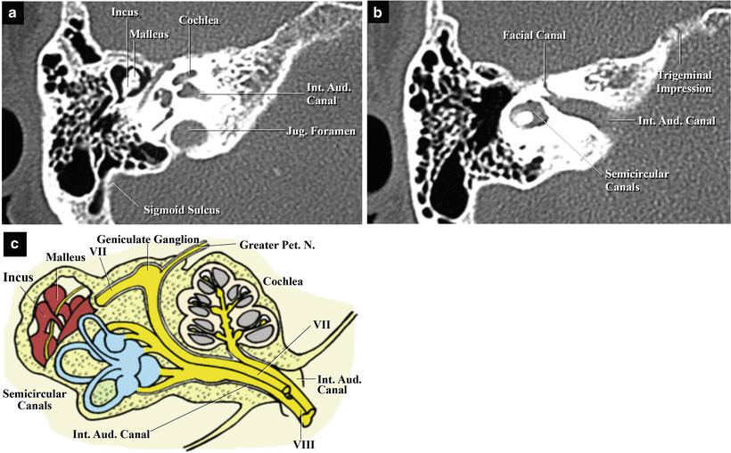 The Temporal Bone Basic Anatomy And Approaches To Internal
 Endoscopic Transmastoid Posterior Petrosal Approach For
 Figure 1 From High Jugular Bulb In The Translabyrinthine
Anatomy Of The Fundus Of The Internal Acoustic Meatus
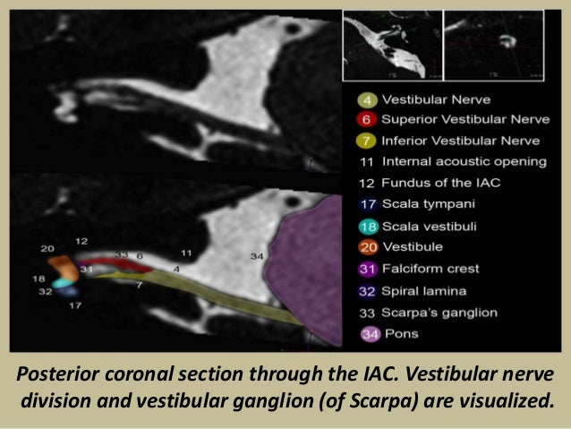 Presentation1 Pptx Radiological Anatomy Of The Petrous Bone
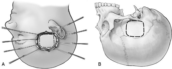 The Middle Fossa Approach Barrow
 Internal Acoustic Canal Radiology Reference Article
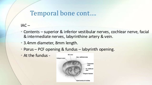
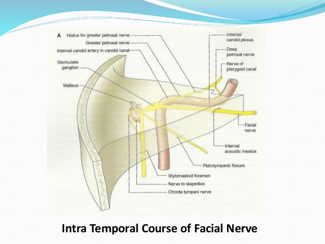
Posting Komentar
Posting Komentar