The epidermis is the outermost layer that provides a protective waterproof seal over the body. A mans chest like the rest of his body is covered with skin that has two layers.

The sternum or breastbone is a long flat bone in the center of the chest.
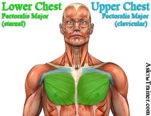
Upper chest anatomy. This thoracic and pulmonary anatomy tool is especially designed for students of anatomy medical and paramedical studies. These important muscles control many motions that involve moving the arms and head such as throwing a ball looking up at the sky and raising your hand. The chest as part of this group enables you to perform pushing actions such as the barbell bench press or a daily activity such as moving a heavy dresser.
One important organ in the chest is the thymus a small butterfly shaped organ located between the heart and the sternum or breastbone. The thorax includes the thoracic cavity and the thoracic wall. The muscles of the chest and upper back occupy the thoracic region of the body inferior to the neck and superior to the abdominal region and include the muscles of the shoulders.
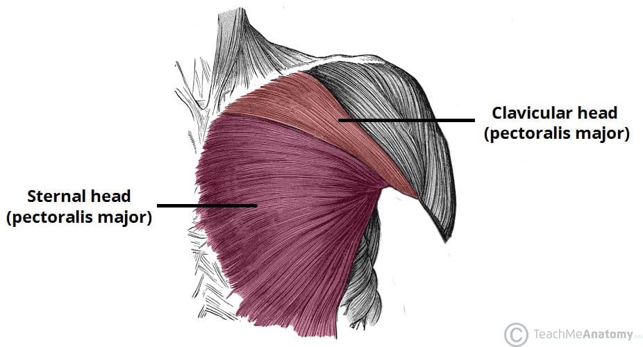
The thorax or chest is a part of the anatomy of humans and various other animals located between the neck and the abdomen. It protects the heart and also serves as the connection point for the costal cartilage. This large fan shaped muscle stretches from the armpit up to the collarbone and down across the lower chest region on both sides of the chest.
The chest is part of a larger group of pushing muscles found in the upper body. This organ belongs to the immune system and its job is to. The diaphragm forms the upper surface of the abdomen.
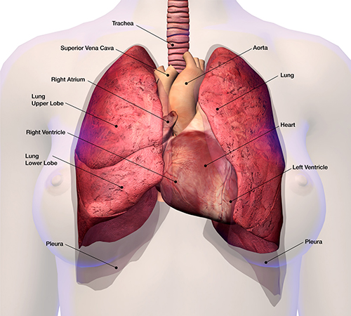
The clavicle or collarbone. Anatomical illustrations this e anatomy module presents an illustrated anatomy of the lungs trachea bronchi pleural cavity and pulmonary vessels. The abdomen commonly called the belly is the body space between the thorax chest and pelvis.
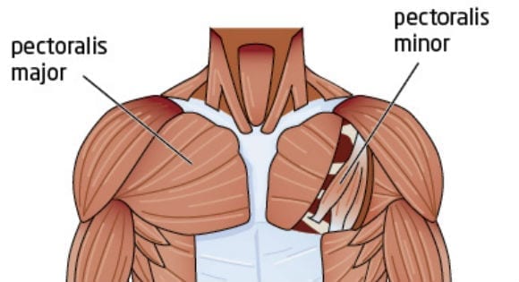
Anatomy of the chest and the lungs. It contains organs including the heart lungs and thymus gland as well as muscles and various other internal structures. The major muscle in the chest is the pectoralis major.
The muscle group that makes up the chest is in the front of the body and includes the sternalis near the sternum the serratus anterior near the armpits the deltoid in the front of the shoulder the pectoralis major at the front center of the chest the pectoralis minor also near the front center of the chest and the external oblique near the lower side of the body. At the level of the pelvic bones the abdomen.
 Atlas Of Surface Anatomy Hadzic S Peripheral Nerve Blocks
 Introduction To Anatomy And Physiology Online Student
Muscles Of The Thoracic Wall Chest Muscles Anatomy
 8 Secrets For Building Your Best Upper Chest T Nation
Best Practices For Treating Diaphragmatic Endometriosis
 Best 5 Exercises For Bigger Chest Fitaitan
 Vascular Anatomy Of The Neck And Upper Thorax Medivisuals
 How To Develop A Man S Pectorals With Strength Training
 Muscles Of The Pectoral Region Major Minor Teachmeanatomy
 Chest Muscle Diagram Chest Muscle Diagram Human Anatomy
 Best Chest Exercises For Men Pecs Anatomy And Chest
 Upper Back Pain From Intercostal Muscle Strain
 Upper Anterior Muscles Anatomy
Do You Suffer With Upper Back Pain Gareth Warburton
What Are The Best Ways To Build Pectoral Muscles
 What Is The Exercise For Upper Chest Muscle Activation Quora
Upper Chest Neck Pain And Tightness
 Chest Workout 5 Exercises To Build The Upper Chest
 Superior Vena Cava Syndrome Cancer Net
 Home Chest Workouts For Upper Lower Pecs With Without
 Anatomy Of Male Muscles In Upper Body Anterior View Photographic Print
 Chest Muscle Injuries Strains And Tears Of The Pectoralis


Posting Komentar
Posting Komentar