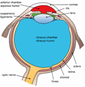
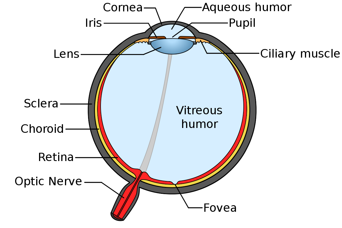
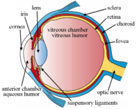
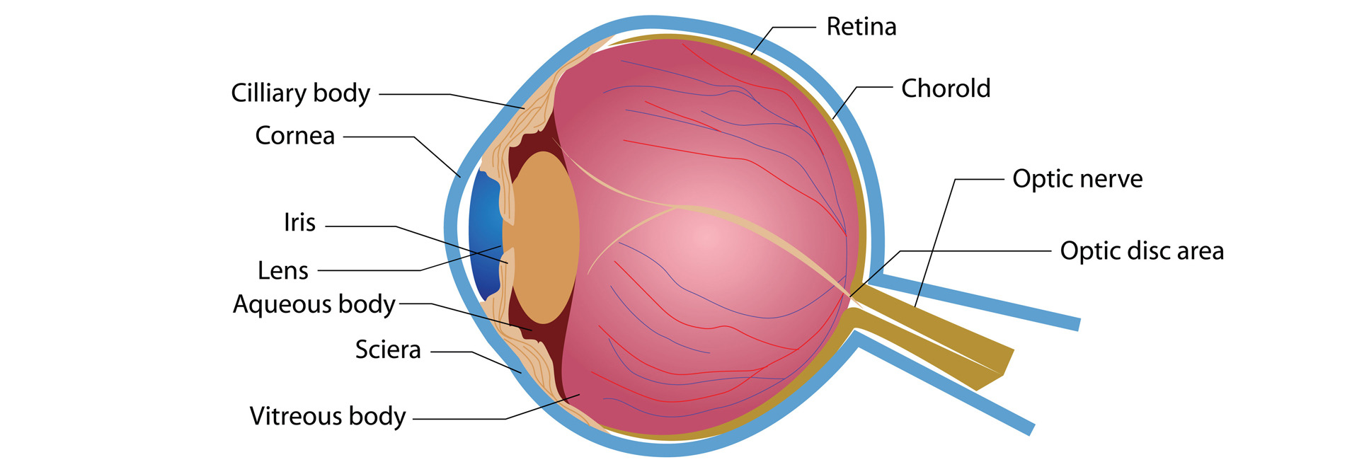
The orbit is the bony eye socket of the skull. Most of the eye is filled with a clear gel called the vitreous.
 Eye Anatomy Micro Chirurgia Oculare
The anterior segment is the first third of the eye from the cornea to the lens.
Eye lens anatomy. The eyelids serve to protect the eye from foreign matter such as dust dirt. Parts of the eye. Lens lens in anatomy a nearly transparent biconvex structure suspended behind the iris of the eye the sole function of which is to focus light rays onto the retina.
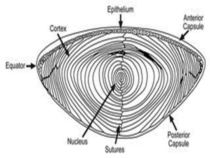
In other words it focuses the light rays that pass through it and onto the retina in order to create clear images of objects that are positioned at various distances. The diagrams below show cross sections of the human eyeball. The iris of the eye functions like the diaphragm of a camera controlling the amount.
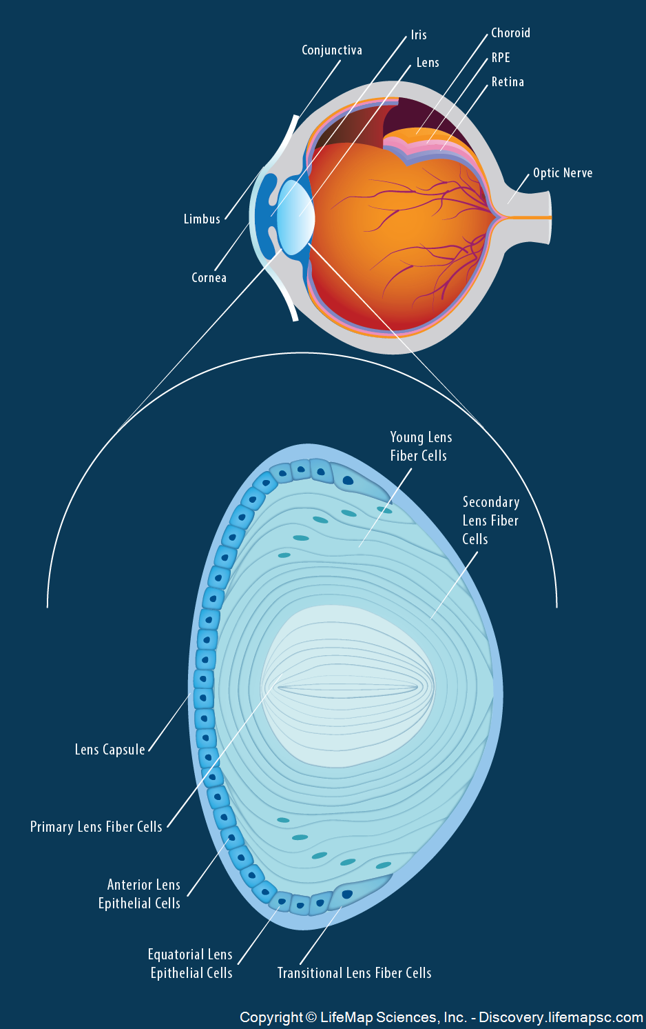
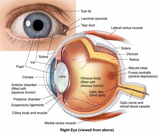
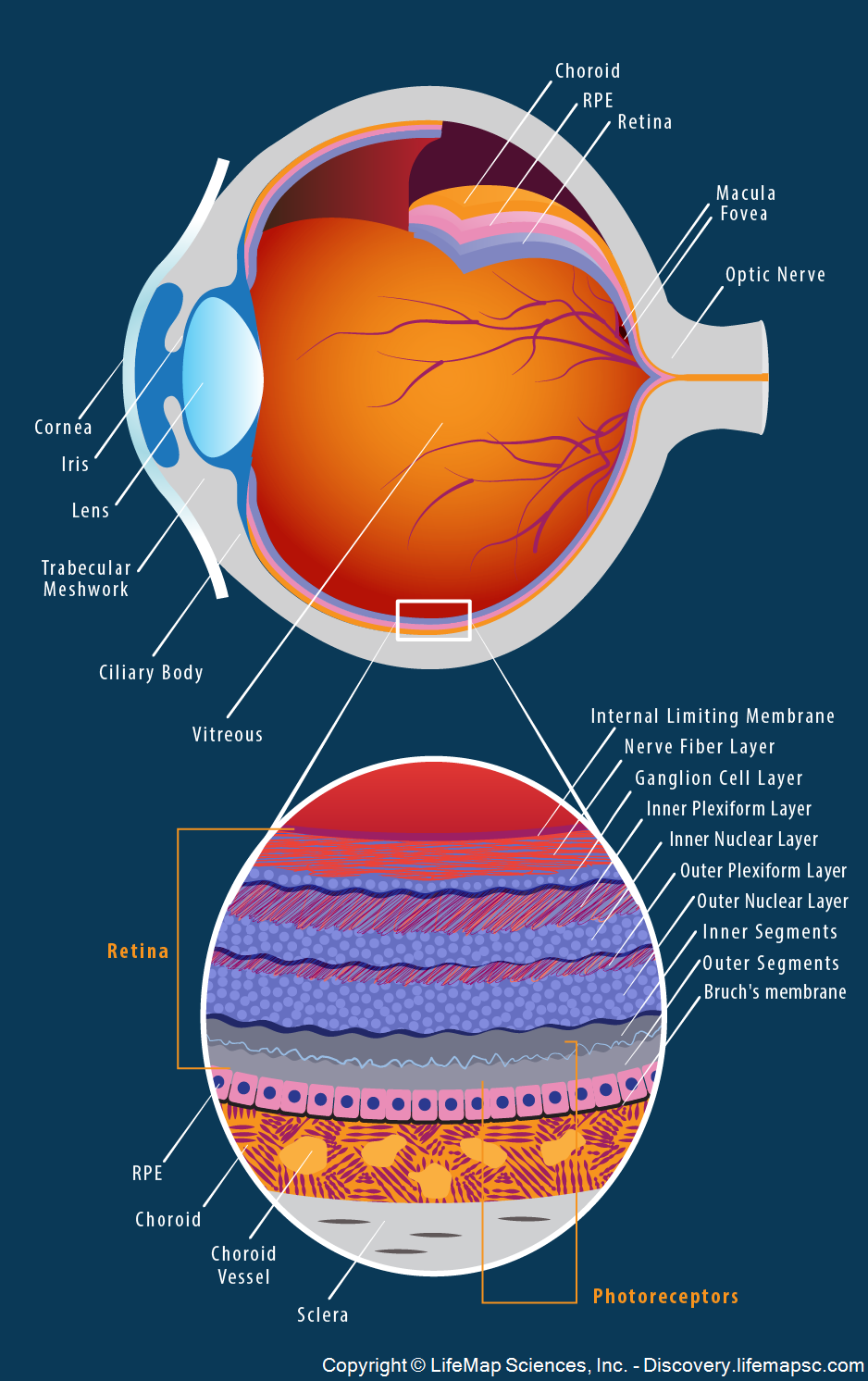
Anatomy of the eye lens the anatomy and structure of the adult human lens infographic in this image you will find limbus cornea conjuctiva iris lens choroid rpe retina optic nerve in eye lens anatomy in detail. Just behind the iris and pupil lies the lens which helps focus light on the back of your eye. Anatomy of the eye.
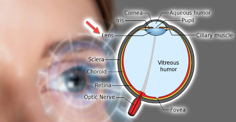
Light projects through your pupil and. This adjustment of the lens is known as accommodation see also below. Anatomy and physiology of the eye eye anatomy facts.
The vitreous cavity lies between the lens and the back of the eye. In a number of ways the human eye works much like a digital camera. The eyes crystalline lens is located directly behind the.
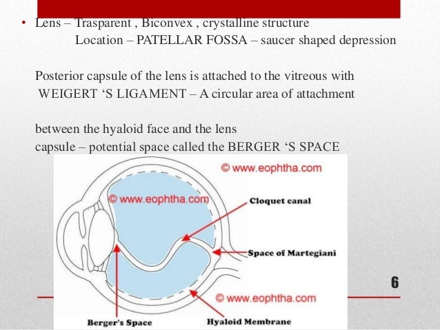
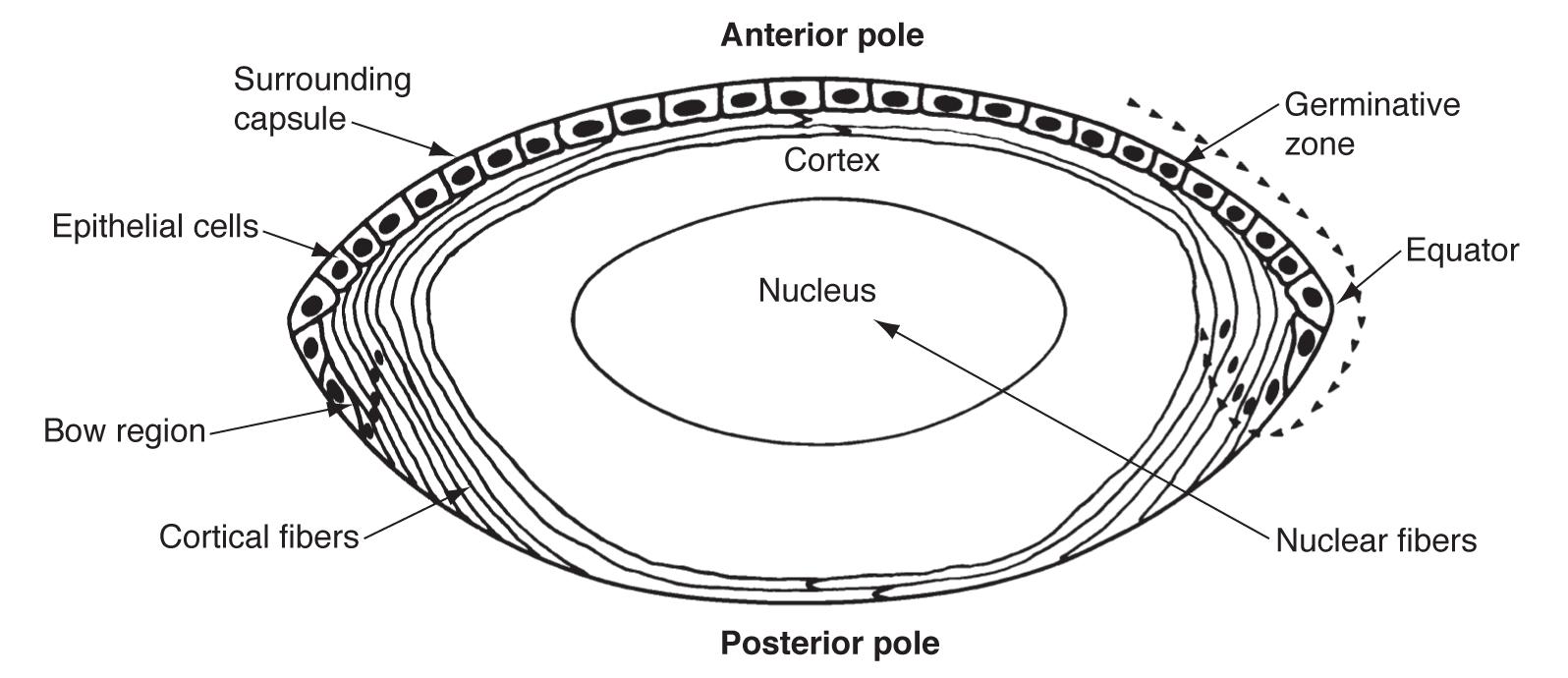
Vision is by far the most used of the five senses and is one. Small fibers called zonules are attached to the capsule holding the lens suspending it from the eye wall. The lens changes shape to help the eye focus on objects up close.
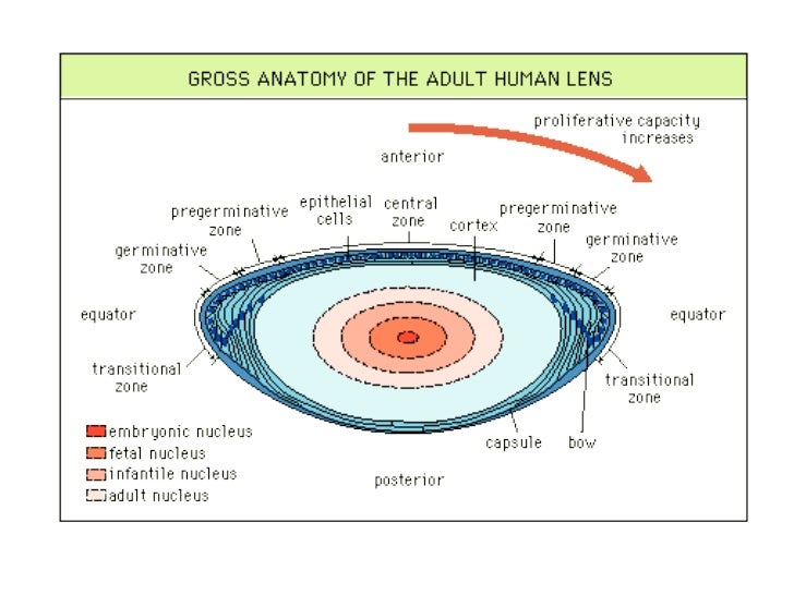
By changing its shape the lens changes the focal distance of the eye. This article explores the anatomy of the eye looking at the different structures of the human eye and their function. You may also find young lens fiber cells secondary lens fiber cells lens capsule primary lens fiber cells in it.
Light is focused primarily by the cornea the clear front surface of the eye. Lens anatomy the lens by changing shape functions to change the focal distance of the eye so that it can focus on objects at various distances thus allowing a sharp real image of the object of interest to be formed on the retina. It also works together with the cornea to refract or bend light.
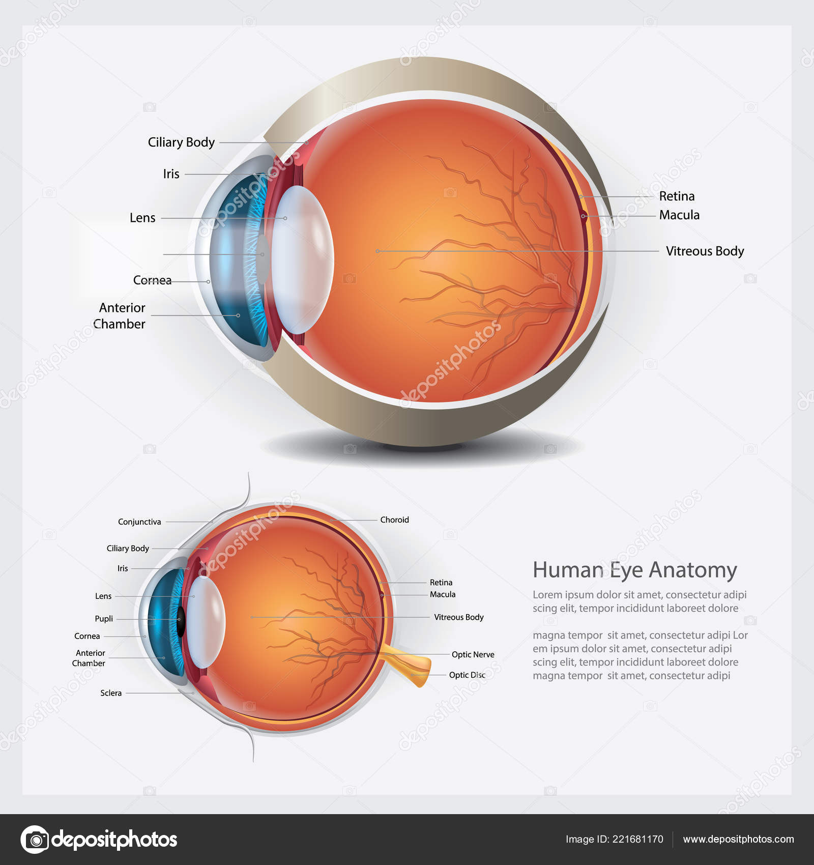
Vision is our window to the outside world. The lens is located in the eye. The anterior segment includes the iris ciliary body and two fluid filled chambers also called anterior and posterior.
The lens is suspended just posteriorly to the iris and divides the anterior and posterior segments of the eye. The lens focuses light toward the back of the eye. The eye is the organ responsible for vision.
 Eye Anatomy Cross Section Of The Lens In 2019 Eye
 Lens Anatomy Wikipedia
 Human Eye Anatomy Normal Lens Vector Illustration Stock
 Lens Anatomy Wikipedia
 Lens Anatomy And Physiology
 Vector Structure Of The Human Eye Anatomy And Medicine Iris
 Optical System Of Eye Gauri S
 Are There Any Parts Of The Human Body That Get Oxygen
 Eye Conditions Florida Eye Clinic
 Anatomy Of The Eye Moorfields Eye Hospital
Lens
 Lens Anatomy Wikipedia

 Anatomy Of The Eye Columbia Eye Clinic

 Eye Lens Anatomy Accommodation Herbal Software
 Vision In Fishes Wikipedia
 Eye Lens Anatomy Images Stock Photos Vectors Shutterstock
Eye Health Anatomy Of The Eye Visionaware
 Lens Anatomy Simple English Wikipedia The Free Encyclopedia

 Ophthalmology Realistic Set Of Contact Lens And Human Eye Anatomy
 Armenian Eyecare Project Anatomy Of The Eye
 Vision And The Eye S Anatomy Healthengine Blog
 Amazon Com Ahawoso Shower Curtain Set With Hooks 72x72


Posting Komentar
Posting Komentar