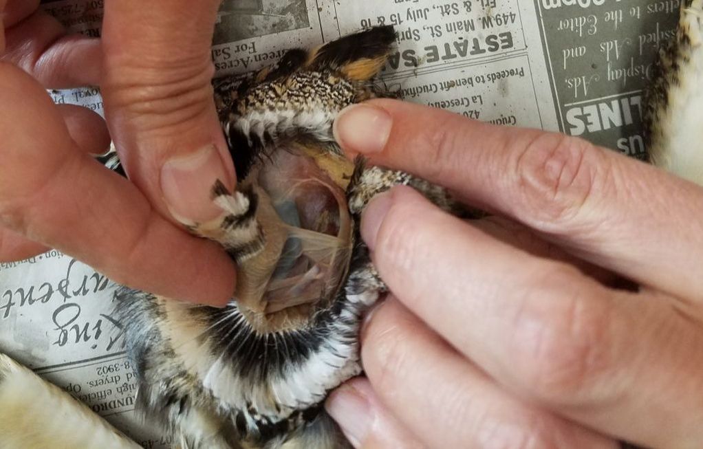
Tear drains from the eyes in to the nose through the tear duct. This is a small tube that runs from the eye to the nasal cavity.
 53 Interesting And Fun Owl Facts Factretriever Com
And the evolution of such large eyes has required a behavioral compromise.

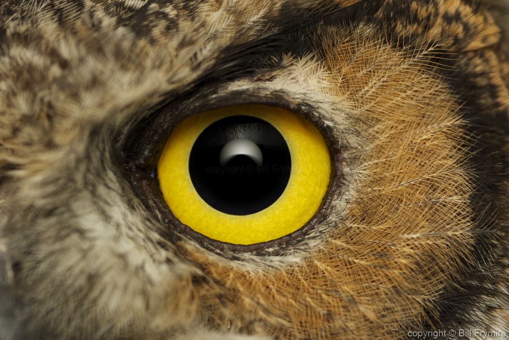
Owl eyes anatomy. An owls eyes are fixed in their sockets so the bird must rotate its neck to look around. O also owls eyes are locked in place and cannot move on their own. Owls see well in daylight too but their color vision is probably very limited.
They also have three eyelids in order to properly protect their eyes. And the evolution of such large eyes has required a behavioral compromise. In the diagram above anatomy of the eye the artery is shown in red while the vein is shown in blue.
Mr is the most sensitive imaging technique for these cord lesions and often provides information that suggests the underlying etiology. An owls eyes are fixed in their sockets so the bird must rotate its neck. This is why a teary eye is usually accompanied by a runny nose.
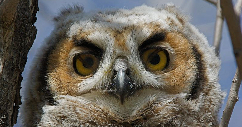
Although the owls eyes sign can be a striking intramedullary finding on mr initially thought indicative of occlusion of the anterior spinal arteries it is a non specific imaging sign and can result from a variety of pathologies including cord infarction cord contusion compressive myelopathy and various infectious or inflammatory conditions. Greater range of sight owls have large eyes placed in the center of the face for greater depth perception. The owl eyes sign also referred to as snake eyes sign or fried eggs sign represents bilaterally symmetric circular to ovoid foci of high t2 weighted signals in the anterior horn cells of the spinal cord and is seen on axial mr imaging.
To protect their eyes owls are equipped with 3 eyelids. They can turn their heads around and even upside down. Owls see well in daylight too but their color vision is probably very limited.
They have a normal upper and lower eyelid the upper closing when the owl blinks and the lower closing up when the owl is asleep. An owls retinal anatomy is similar to that of cats which rival owls in seeing in dim light. It is quite an amazing ability of their anatomy that fascinates people.
Owls actually have a field of vision of about 110 degrees with about 70 degrees of it being binocular vision enabled. An owls retinal anatomy is similar to that of cats which rival owls in seeing in dim light. This cleans and protects the surface of the eye.
The third eyelid is called a nictitating membrane and is a thin layer of tissue that closes diagonally across the eye from the inside to the outside. Therefore the owl must turn its head in order to see whats around it. O greater depth perception allows owls to see more at a greater distance than other birds.
 Owl Facts For Kids Cool Kid Facts
 Snowy Owl With Labels
 Snowy Owl Printout Enchantedlearning Com

 Bird S Eye View National Geographic Society
 Anatomy Of The Eye Human Eye Anatomy Owlcation
 Owl Wikipedia
 Secrets Of The Snowy Owl Habitat Adaptations And Other Facts
 Barn Owls Eyes Diagram Reading Industrial Wiring Diagrams
 544 817 593 Eagle Owl Head

 How To Draw An Owl

 Spotlight On Tawny Owls St Nicks
 Owl Eyes Sign Spinal Cord Radiology Reference Article
 How To Draw An Owl

Owls Study Guide
 How To Draw Owl Eyes Draw An Owl Face Step By Step Birds
 Owl Eyes Vision The Owl Pages
 Pecten Oculi Wikipedia
 The Great Gray Owl Probably Has Bigger Eyes Than You Audubon
Eagle Eyes Raptor Resource Project
 Beautifully Designed Owl Eyes Drawings Page 2 Of 4 Fine
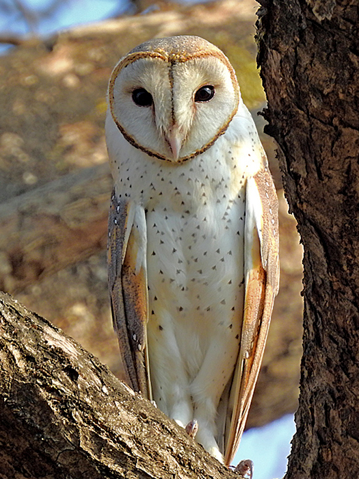
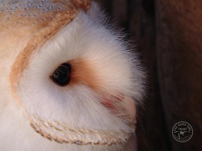 Barn Owl Adaptations The Barn Owl Trust


Posting Komentar
Posting Komentar