19 public playlist includes this case. Spin echo t1 and proton density with fat saturation sequences.
 Mri Anatomy Of The Shoulder Ppt Video Online Download
Knee shoulder shoulder arthrogram ankle elbow wrist hip.
Mri shoulder anatomy. T2 star gradient recall echo images are employed in the assessment of the labrum and for detection of substances that produce susceptibility effects such as calcium hydroxyapatite or loose surgical hardware. Normal shoulder mri for reference. If you continue browsing the site you agree to the use of cookies on this website.
Muhammad bin zulfiqar slideshare uses cookies to improve functionality and performance and to provide you with relevant advertising. Mr is the best imaging modality to examen patients with shoulder pain and instability. Magnetic resonance imaging.
An mri scanner uses a high powered magnet and a computer to create high resolution images of the shoulder and surrounding structures. This mri shoulder coronal cross sectional anatomy tool is absolutely free to use. In part iii we will focus on impingement and rotator cuff tears.
Mri shoulder protocols typically involve fat saturated proton density images that are sensitive to internal derangement. Atlas of shoulder mri anatomy. In shoulder mr part i we will focus on the normal anatomy and the many anatomical variants that may simulate pathology.
Mri of shoulder anatomy dr. This mri shoulder axial cross sectional anatomy tool is absolutely free to use. An mri of the shoulder of a healthy subject was performed in the 3 planes of space coronal axial sagittal commonly used in osteoarticular imagery with two weightings most commonly used to explore the musculo skeletal pathology of the shoulder.
In part ii we will discuss shoulder instability. Well actually there is thickening of the inferior glenohumeral ligament suggesting multidirectional instability but it is still a good study to observe normal anatomy. Use the mouse scroll wheel to move the images up and down alternatively use the tiny arrows on both side of the image to move the images.
Use the mouse scroll wheel to move the images up and down alternatively use the tiny arrows on both side of the image to move the images on both side of the image to move the images. Use the mouse to scroll or the arrows. This webpage presents the anatomical structures found on shoulder mri.
Click on a link to get t1 axial view t2 fatsat axial view t1 coronal view t2 fatsat coronal view t2 fatsat sagittal view.
 Shoulder Mri Radtechonduty

 Posterior Labral Tear Shoulder Elbow Orthobullets
 Mri Shoulder Anatomy Shoulder Coronal Anatomy Free Cross
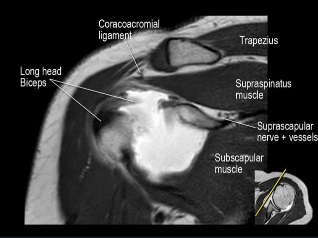
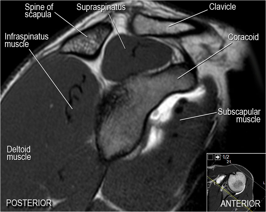
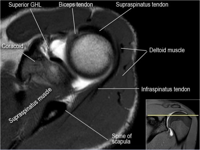
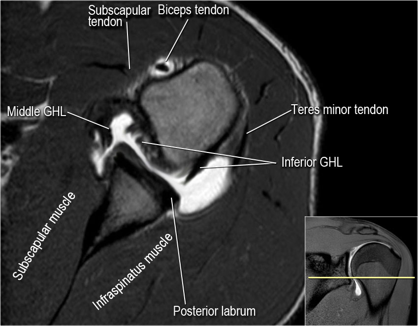 The Radiology Assistant Shoulder Mr Anatomy
 Musculoskeletal Mri
![]() Medical Imaging And Radiological Anatomy X Ray Ct Mri
Medical Imaging And Radiological Anatomy X Ray Ct Mri
Mri Of Shoulder Anatomy

 Mri Of Shoulder Radiology Lecture Slides Docsity


 Shoulder Impingement 3 Keys To Assessment And Treatment
 Rotator Cuff Radiology Reference Article Radiopaedia Org
 Shoulder Mri Radiographical And Illustrated Anatomical Atlas
 Cables Crescents And Suspension Bridges The Unique Anatomy

 The Radiology Assistant Shoulder Mr Anatomy
 Shoulder Anatomy Shoulder Injuries Chicago Westchester
 Normal And Variant Anatomy Of The Shoulder On Mri
 Shoulder Anatomy Mri Shoulder Axial Anatomy Free Cross
 Mri Shoulder Anatomy Shoulder Coronal Anatomy Free Cross

![]() Mri Arthroscopy Correlation For Shoulder Anatomy And
Mri Arthroscopy Correlation For Shoulder Anatomy And

 Improve Msk Imaging Outcomes Imaging Technology News
 Mri Of Shoulder Anatomy
 Mri Shoulder Anatomy Shoulder Coronal Anatomy Free Cross


Posting Komentar
Posting Komentar