Orbital cavity the bony cavity in the skull containing the eyeball. A long sensory root from the nasociliary branch of v1 10 12 mm with fibers from cornea iris and ciliary body.
 Naso Orbital Ethmoid Fractures Springerlink
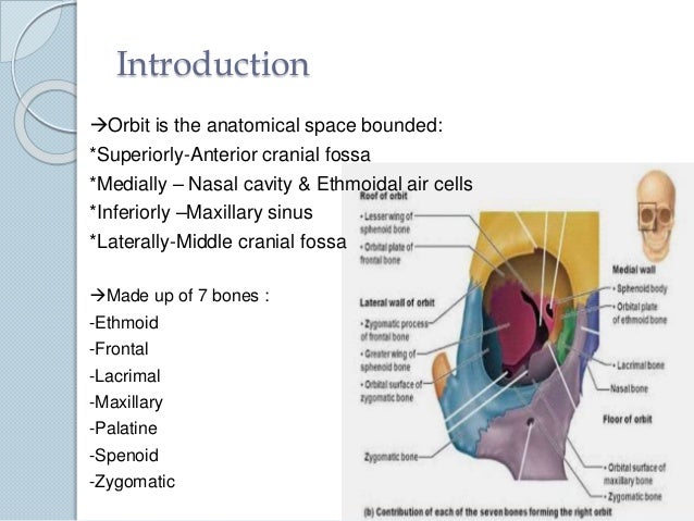
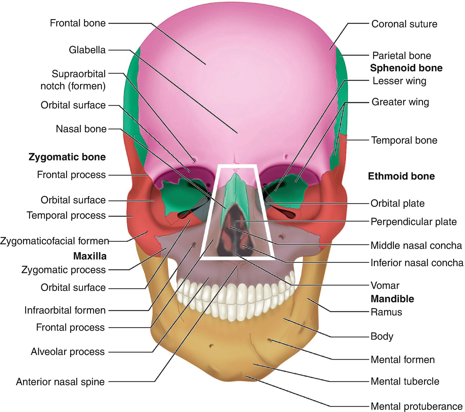
It is formed of parts of the frontal maxillary sphenoid lacrimal zygomatic ethmoid and palatine bones.
Orbital anatomy definition. In the lateral part in the neurocranium of the orbital region two pairs of foramina occur. Bodily cavity cavum cavity anatomy a natural hollow or sinus within the body. Lacrimal bone small fragile bone making up part of the front inner walls of each eye socket and providing room for the passage of the lacrimal ducts.
Orbita ta orbital cavity. Among their topics are the structural and functional anatomy of the orbital region requirements for surgery and basic operative techniques ptosis operation in combination with upper lid blepharoplasty basic principles of ablative fractional. Protects the body keeps harmful material out regulates body temperature senses and responds to the environment and creates important chemicals.
In the adult human the volume of the orbit is 30 millilitres 106 imp fl oz. In anatomy pertaining to the orbit the bony cavity that contains the eyeball. Sympathetic root comes from ica plexus and enters through superior orbital fissure.
Short motor root from inferior division of cn iii supplying the inferior oblique postganglionic fibers supply the iris sphincter. The medial orbital rim is less defined than the other rims. Orbit anatomy in anatomy the orbit is the cavity or socket of the skull in which the eye and its appendages are situated.
Cranial orbit eye socket orbit. Orbit definition the curved path usually elliptical described by a planet satellite spaceship etc around a celestial body as the sun. The bony cavity containing the eyeball and its adnexa.
Of or relating to an orbit. 101 us fl oz. Orbit can refer to the bony socket or it can also be used to imply the contents.
The electron in that state. The entire wall is thin from the base to the apex but it is strengthened by the perpendicular septa of the ethmoid sinus. The medial orbital walls are parallel to the sagittal plane and have the greatest degree of superioinferior curvature.
A wave function describing the state of a single electron in an atom atomic orbital or in a molecule molecular orbital. Anatomy and physiology chapter 1.
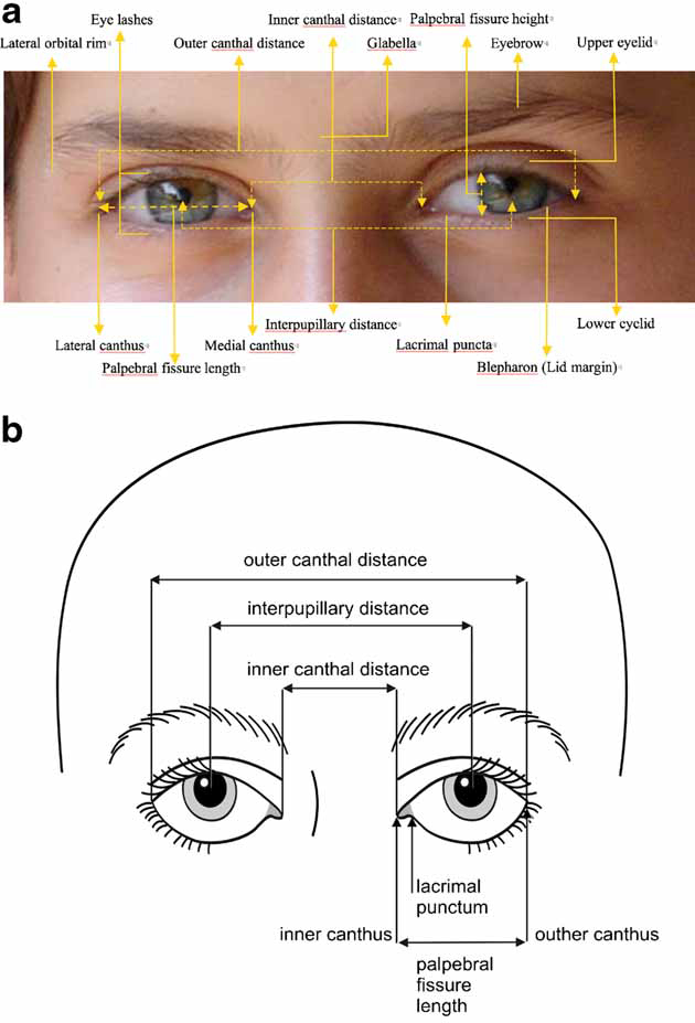 Elements Of Morphology Human Malformation Terminology
 Figure 4 From Use Of High Resolution Microscopy Coil Mri For
 Bony Orbit Radiology Reference Article Radiopaedia Org
 Ch 2 Surface Anatomy At Florida Southwestern State College
 Ocular Adnexa Definition Anatomy Study Com
 Orbital Cellulitis Intechopen
 Multimodal Treatment Of Orbital Tumors Neupsy Key
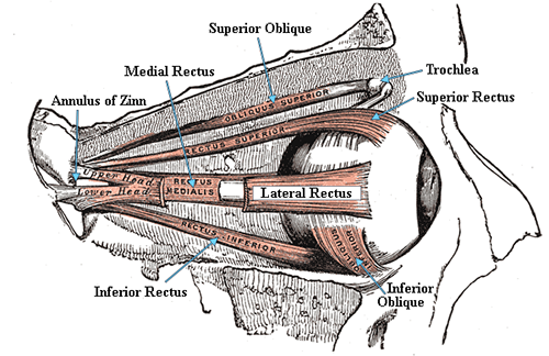 Extraocular Muscles And Movements
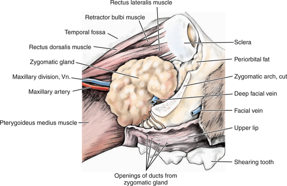 Orbit Veterian Key
 Orbital Movements Of Axis Definition Of Orbital Movements
 Superior Orbital Fissure Wikipedia
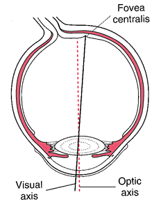
Orbital Axis
Superior Orbital Fissure
 Transcranial Approach To The Orbit Microsurgical Anatomy In
 Endoscopic Surgery Of The Orbit Anatomy Pathology And
 Endoscopic Surgery Of The Orbit Anatomy Pathology And

 Definition Of Ethmoid Bone In The Medical Dictionary By The
 Ophthalmic Emergency Orbital Cellulitis
 Anatomy Of The Left Orbital Apex Highlighting The
 Midface Authors Added Material Ao Surgery Reference
 Orbit Anatomy
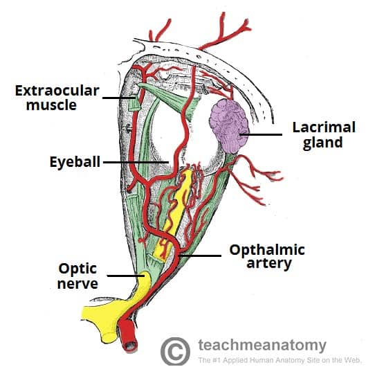
The Bony Orbit Borders Contents Fractures Teachmeanatomy
 The Radiology Assistant Orbita Pathology
 Midfacial Fractures Springerlink


Posting Komentar
Posting Komentar