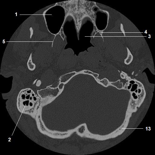
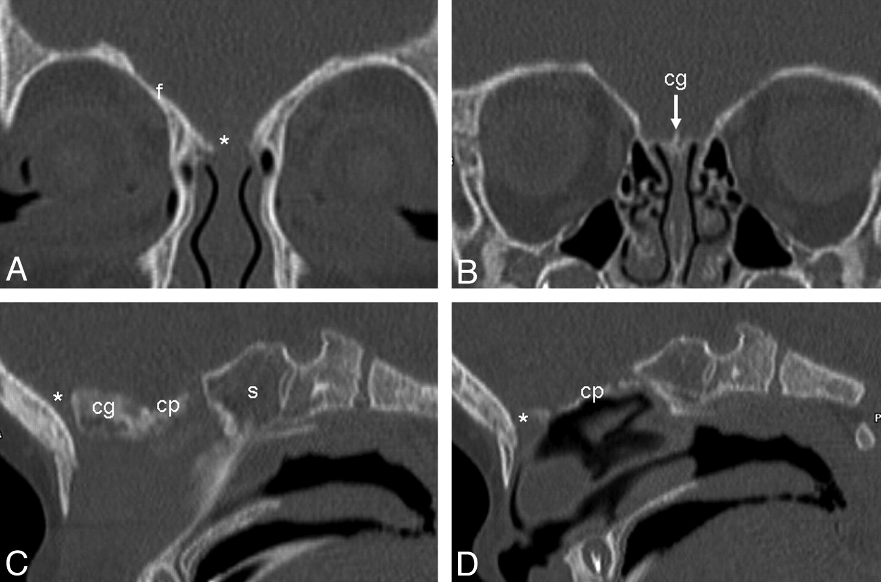
2 superior orbital fissure. Skull ct anatomy the sagittal suture is the line where the right and left parietal bone are in contact.
Detailed anatomy enter this module for a more detailed review of skull base anatomy.

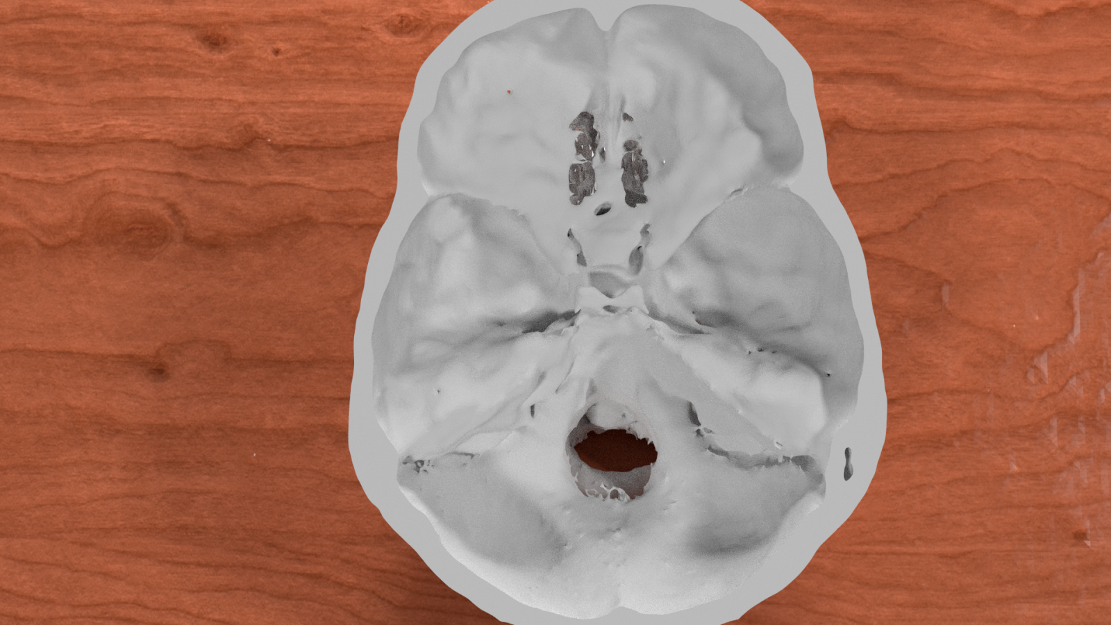
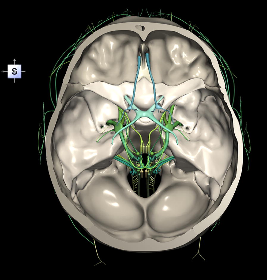
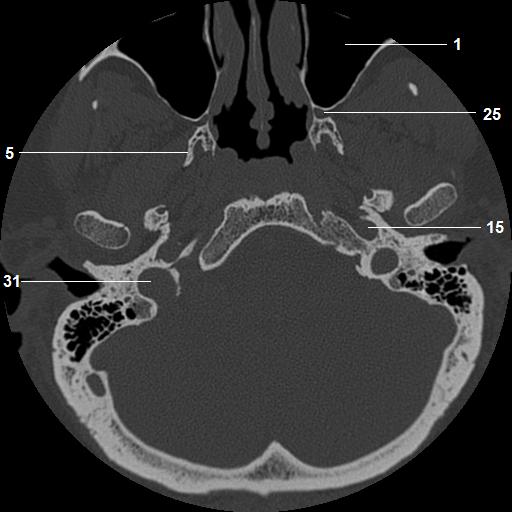
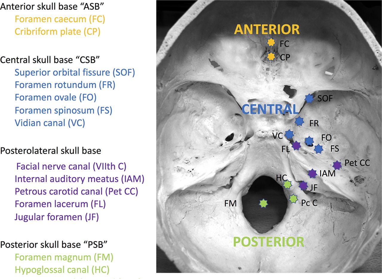
Skull base anatomy ct. Navigating the skull base identify the petro occipital fissure to navigate the major structures of the skull base. Basic skull base anatomy. A axial three dimensional reconstructed ct image with color coded overlay shows the skull base sections.
3 anterior clinoid process. The coronal suture is the line where the parietal bone frontal bone and are in contact. The module interface is meant to mimic a radiology workstation with adjacent image scrolling via arrow keys and or mouse wheel button.
Given that the file is large loading may take a few minutes. To load the skull base ct anatomy module in a new window click on its image above. Blue temporal bones fuchsia nasal bones green ethmoid bone light pink vomer purple occipital bones teal sphenoid bone yellow zygomatic bones.
Basic anatomy review the bones sutures and fissures that comprise the skull base. Ct anatomy of skull base. Blue central skull base csb purple posterior skull base teal anterior skull base asb.
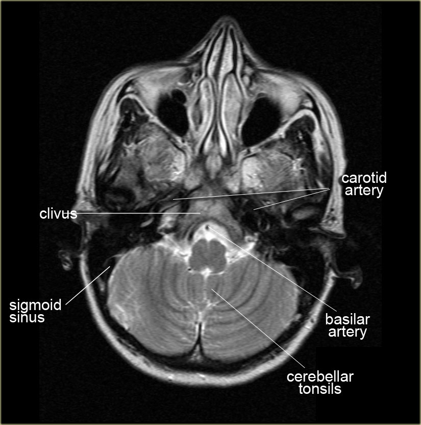
B axial ct image with color coded overlay shows the skull base bones. The skull base can be evaluated by computed tomography ct which will demonstrate the bony structures of the skull base with its foramina and fissures for vessels and cranial nerves the temporal bone and sinonasal cavities.

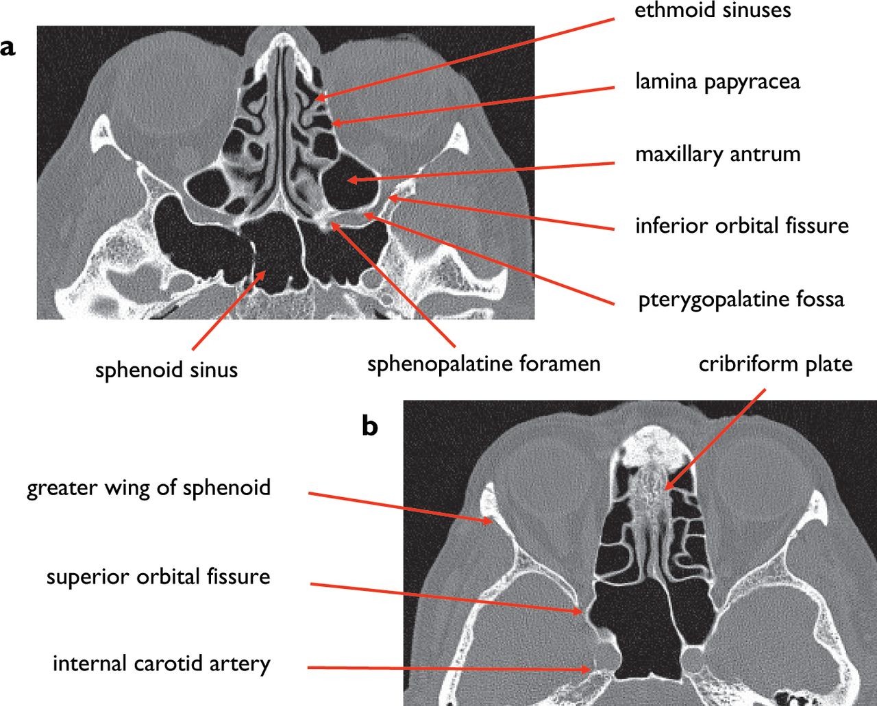
 Imaging Of Paranasal Sinuses And Anterior Skull Base And

 Skull Base Petrous Apex Tumors Background History Of The
 The Radiology Assistant Brain Anatomy
 3d Printed Skull Base Generated From Ct Scan Data Accurately
 A 3d Stereotactic Atlas Of The Adult Human Skull Base

 Middle Skull Base Plastic Surgery Key

 Ecr 2015 C 0264 Imaging Of The Anterior And Central
 Normal Anatomy Of The Base Of The Skull Orbit Pituitary
 A 3d Stereotactic Atlas Of The Adult Human Skull Base
 Skull Base Tumors Uci Head And Neck Surgery Uci Ent
 Headneckbrainspine

Anatomy And Pathology Of The Skull Base Ct And Mri Imaging
 Skull Base Imaging Anatomy Pathology And Protocols

 Ao Surgery Reference
 Axial Ct Bone Window Of Skull Base From Inferior To Superior
Ct Of Normal Developmental And Variant Anatomy Of The
 Skull Base Imaging Anatomy Pathology And Protocols
Ct And Mr Imaging Of The Central Skull Base Part 2

 The Anatomy Of The Human Skull Download Free 3d Model By
 Skull Base Ct And Mr Anatomy Module Tutorial
 Base Of Skull Wikipedia
 Normal Anatomy Of The Base Of The Skull Orbit Pituitary


Posting Komentar
Posting Komentar